Office of Governor Dan Malloy|sourceurl=https://flickr.com/photos/57939039@N08/43757681472%7Carchive=%7Creviewdate=2021-11-22 00:57:16|reviewlicense=cc-by-2.0|reviewer=FlickreviewR 2
Wikimedia Commons
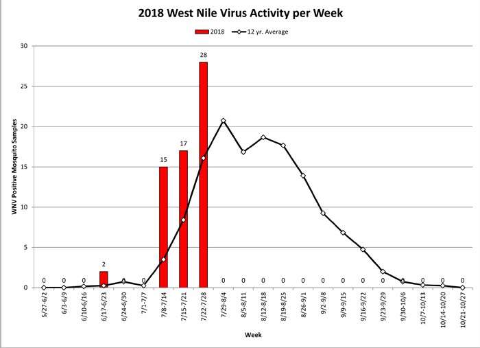
Description: Thursday, August 2, 2018 -- Governor Dannel P. Malloy and state public health officials are advising Connecticut residents to be aware of a rapid build-up of West Nile virus (WNV) activity within the state as recent tests show that infected mosquitoes are rising at levels higher than normal for this time of year. The Connecticut Agricultural Experiment Station (CAES) has detected WNV-infected mosquitoes in 19 municipalities this year, including Bethany, Bridgeport, Darien, East Haven, Easton, Franklin, Greenwich, Hartford, Madison, Manchester, Meriden, New Canaan, New Haven, Stamford, Stratford, Waterbury, Waterford, West Haven, and Weston. Date: 2 August 2018, 12:10. Source: Gov. Malloy and Public Health Officials Advise Residents of Increased West Nile Virus Activity This Season. Author: Dannel Malloy.
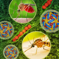
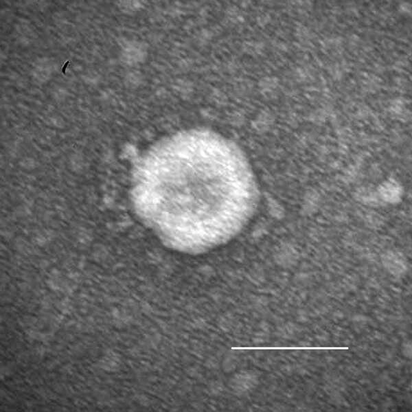
Summary.mw-parser-output table.commons-file-information-table,.mw-parser-output.fileinfotpl-type-information{border:1px solid #a2a9b1;background-color:#f8f9fa;padding:5px;font-size:95%;border-spacing:2px;box-sizing:border-box;margin:0;width:100%}.mw-parser-output table.commons-file-information-table>tbody>tr,.mw-parser-output.fileinfotpl-type-information>tbody>tr{vertical-align:top}.mw-parser-output table.commons-file-information-table>tbody>tr>td,.mw-parser-output table.commons-file-information-table>tbody>tr>th,.mw-parser-output.fileinfotpl-type-information>tbody>tr>td,.mw-parser-output.fileinfotpl-type-information>tbody>tr>th{padding:4px}.mw-parser-output.fileinfo-paramfield{background:#ccf;text-align:right;padding-right:0.4em;width:15%;font-weight:bold}.mw-parser-output.commons-file-information-table+table.commons-file-information-table,.mw-parser-output.commons-file-information-table+div.commons-file-information-table>table{border-top:0;padding-top:0;margin-top:-8px}@media only screen and (max-width:719px){.mw-parser-output table.commons-file-information-table,.mw-parser-output.commons-file-information-table.fileinfotpl-type-information{border-spacing:0;padding:0;word-break:break-word;width:100%!important}.mw-parser-output.commons-file-information-table>tbody,.mw-parser-output.fileinfotpl-type-information>tbody{display:block}.mw-parser-output.commons-file-information-table>tbody>tr>td,.mw-parser-output.commons-file-information-table>tbody>tr>th,.mw-parser-output.fileinfotpl-type-information>tbody>tr>td,.mw-parser-output.fileinfotpl-type-information>tbody>tr>th{padding:0.2em 0.4em;text-align:left;text-align:start}.mw-parser-output.commons-file-information-table>tbody>tr,.mw-parser-output.fileinfotpl-type-information>tbody>tr{display:flex;flex-direction:column}.mw-parser-output.commons-file-information-table+table.commons-file-information-table,.mw-parser-output.commons-file-information-table+div.commons-file-information-table>table{margin-top:-1px}.mw-parser-output.fileinfo-paramfield{box-sizing:border-box;flex:1 0 100%;width:100%}} Description: Artwork featuring female Culex quinquefasciatus mosquitoes—which transmit West Nile virus (images courtesy of CDC), a cryo-EM reconstruction of West Nile virus (courtesy of NIH 3D Print Exchange), and a transmission electron micrograph of West Nile virus particles (orange) replicating within the cytoplasm of an infected VERO E6 cell (green), captured at the NIAID Integrated Research Facility (IRF) in Fort Detrick, Maryland. Credit: NIAID and CDC. Date: 10 August 2022, 12:39. Source: West Nile Virus. Author: NIAID.
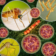
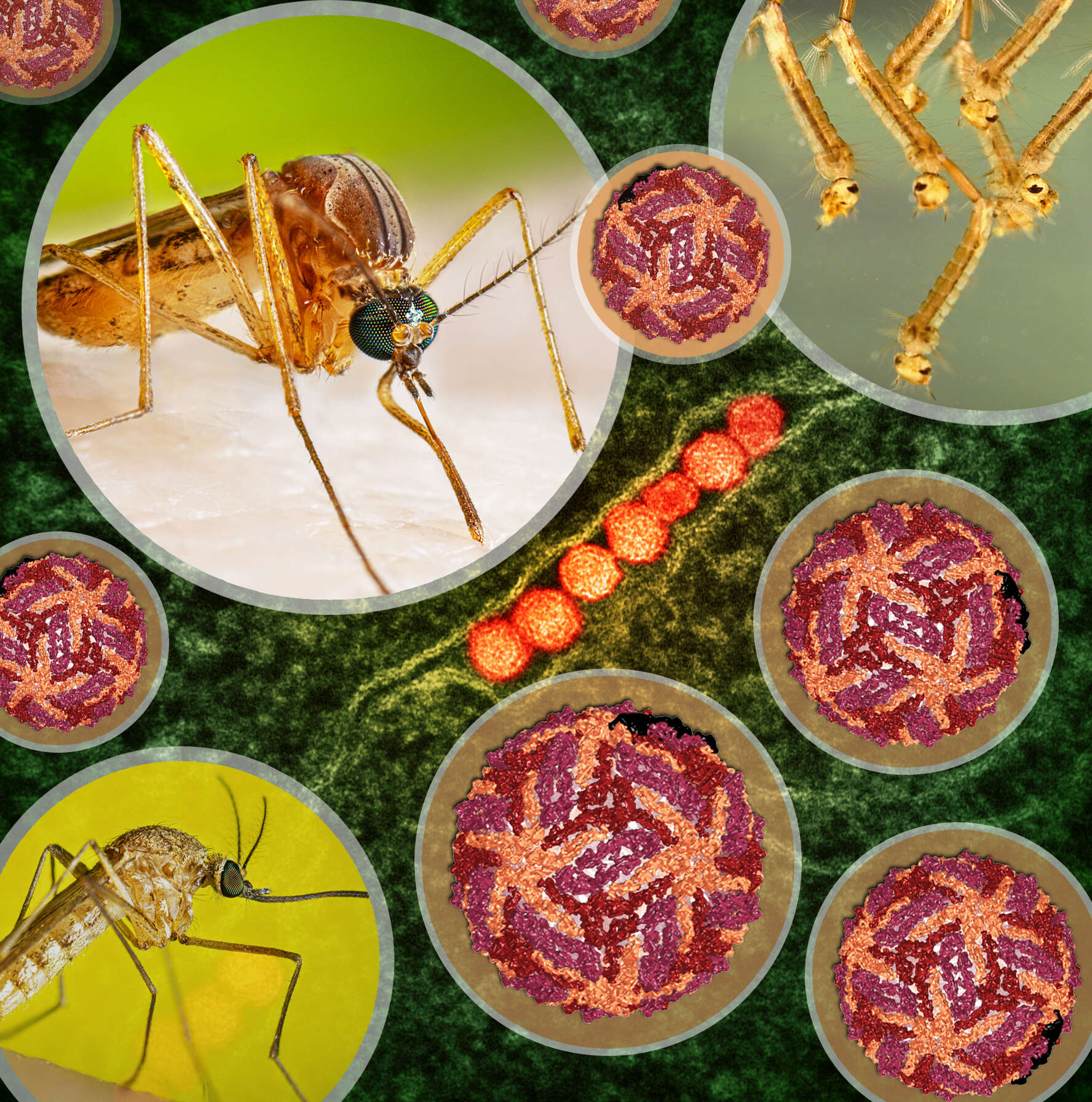
Summary.mw-parser-output table.commons-file-information-table,.mw-parser-output.fileinfotpl-type-information{border:1px solid #a2a9b1;background-color:#f8f9fa;padding:5px;font-size:95%;border-spacing:2px;box-sizing:border-box;margin:0;width:100%}.mw-parser-output table.commons-file-information-table>tbody>tr,.mw-parser-output.fileinfotpl-type-information>tbody>tr{vertical-align:top}.mw-parser-output table.commons-file-information-table>tbody>tr>td,.mw-parser-output table.commons-file-information-table>tbody>tr>th,.mw-parser-output.fileinfotpl-type-information>tbody>tr>td,.mw-parser-output.fileinfotpl-type-information>tbody>tr>th{padding:4px}.mw-parser-output.fileinfo-paramfield{background:#ccf;text-align:right;padding-right:0.4em;width:15%;font-weight:bold}.mw-parser-output.commons-file-information-table+table.commons-file-information-table,.mw-parser-output.commons-file-information-table+div.commons-file-information-table>table{border-top:0;padding-top:0;margin-top:-8px}@media only screen and (max-width:719px){.mw-parser-output table.commons-file-information-table,.mw-parser-output.commons-file-information-table.fileinfotpl-type-information{border-spacing:0;padding:0;word-break:break-word;width:100%!important}.mw-parser-output.commons-file-information-table>tbody,.mw-parser-output.fileinfotpl-type-information>tbody{display:block}.mw-parser-output.commons-file-information-table>tbody>tr>td,.mw-parser-output.commons-file-information-table>tbody>tr>th,.mw-parser-output.fileinfotpl-type-information>tbody>tr>td,.mw-parser-output.fileinfotpl-type-information>tbody>tr>th{padding:0.2em 0.4em;text-align:left;text-align:start}.mw-parser-output.commons-file-information-table>tbody>tr,.mw-parser-output.fileinfotpl-type-information>tbody>tr{display:flex;flex-direction:column}.mw-parser-output.commons-file-information-table+table.commons-file-information-table,.mw-parser-output.commons-file-information-table+div.commons-file-information-table>table{margin-top:-1px}.mw-parser-output.fileinfo-paramfield{box-sizing:border-box;flex:1 0 100%;width:100%}} Description: Artwork featuring female Culex quinquefasciatus mosquitoes—which transmit West Nile virus (images courtesy of CDC), Culex mosquito larvae(image courtesy of CDC), a cryo-EM reconstruction of West Nile virus (courtesy of NIH 3D Print Exchange), and a transmission electron micrograph of West Nile virus particles (orange) replicating within the cytoplasm of an infected VERO E6 cell (green), captured at the NIAID Integrated Research Facility (IRF) in Fort Detrick, Maryland. Credit: NIAID and CDC. Date: 11 August 2022, 10:50. Source: West Nile Virus. Author: NIAID.
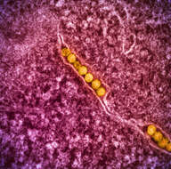
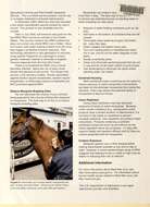
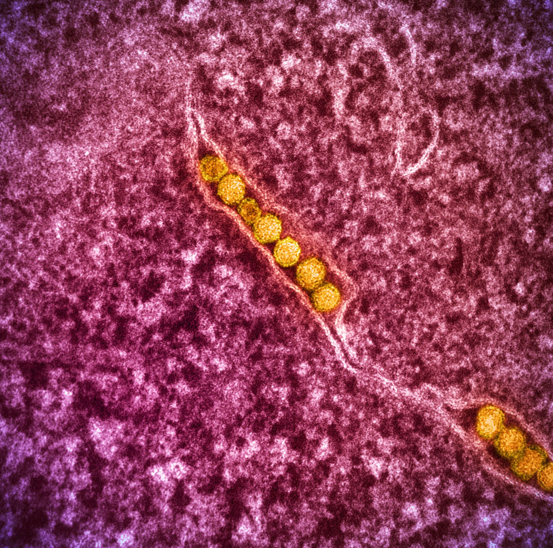
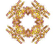
Summary.mw-parser-output table.commons-file-information-table,.mw-parser-output.fileinfotpl-type-information{border:1px solid #a2a9b1;background-color:#f8f9fa;padding:5px;font-size:95%;border-spacing:2px;box-sizing:border-box;margin:0;width:100%}.mw-parser-output table.commons-file-information-table>tbody>tr,.mw-parser-output.fileinfotpl-type-information>tbody>tr{vertical-align:top}.mw-parser-output table.commons-file-information-table>tbody>tr>td,.mw-parser-output table.commons-file-information-table>tbody>tr>th,.mw-parser-output.fileinfotpl-type-information>tbody>tr>td,.mw-parser-output.fileinfotpl-type-information>tbody>tr>th{padding:4px}.mw-parser-output.fileinfo-paramfield{background:#ccf;text-align:right;padding-right:0.4em;width:15%;font-weight:bold}.mw-parser-output.commons-file-information-table+table.commons-file-information-table,.mw-parser-output.commons-file-information-table+div.commons-file-information-table>table{border-top:0;padding-top:0;margin-top:-8px}@media only screen and (max-width:719px){.mw-parser-output table.commons-file-information-table,.mw-parser-output.commons-file-information-table.fileinfotpl-type-information{border-spacing:0;padding:0;word-break:break-word;width:100%!important}.mw-parser-output.commons-file-information-table>tbody,.mw-parser-output.fileinfotpl-type-information>tbody{display:block}.mw-parser-output.commons-file-information-table>tbody>tr>td,.mw-parser-output.commons-file-information-table>tbody>tr>th,.mw-parser-output.fileinfotpl-type-information>tbody>tr>td,.mw-parser-output.fileinfotpl-type-information>tbody>tr>th{padding:0.2em 0.4em;text-align:left;text-align:start}.mw-parser-output.commons-file-information-table>tbody>tr,.mw-parser-output.fileinfotpl-type-information>tbody>tr{display:flex;flex-direction:column}.mw-parser-output.commons-file-information-table+table.commons-file-information-table,.mw-parser-output.commons-file-information-table+div.commons-file-information-table>table{margin-top:-1px}.mw-parser-output.fileinfo-paramfield{box-sizing:border-box;flex:1 0 100%;width:100%}} Description: Transmission electron micrograph of West Nile virus particles (gold) replicating within the cytoplasm of an infected VERO E6 cell (pink). Image captured at the NIAID Integrated Research Facility (IRF) in Fort Detrick, Maryland. Credit: NIAID. Date: 8 August 2022, 12:58. Source: West Nile Virus. Author: NIAID.
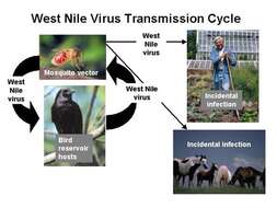
Description: English: Flowchart showing the West Nile virus transmission cycle. Date: 6 September 2012, 16:17:42. Source: https://www.cdc.gov/ncidod/dvbid/westnile/cycle.htm. Author: Centers for Disease Control and Prevention.
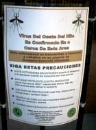
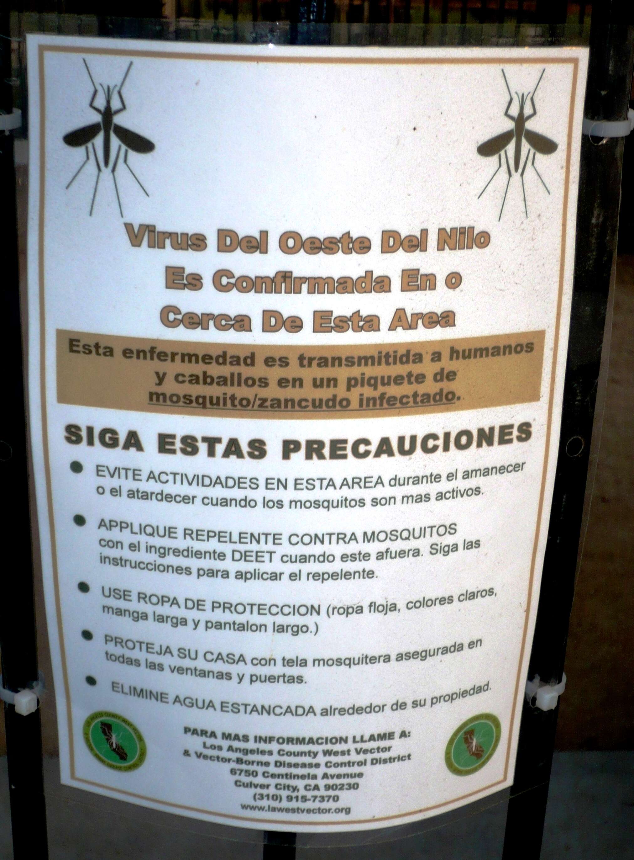
Description: Warning in Spanish for West Nile Virus, Playa Vista, Los Angeles. March 2008. Text of sign translated to English: West Nile Virus Confirmed In or Near This Area. This disease is transmitted to humans and horses from bites by infected mosquitoes. Follow These Precautions: - Avoid activities in this area during dawn or dusk, when mosquitoes are most active. - Apply mosquito repellent containing DEET when outdoors. Follow the instructions to apply repellent. - Wear protective clothing (loose clothing, light colors, long sleeves, and long pants.) - Protect your home with mosquito net fabric around all windows and doors. - Eliminate stagnant water around your property. Date: March 2008. Source: Own work. Author: Polylerus.
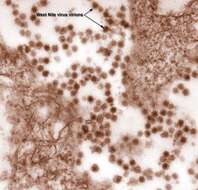
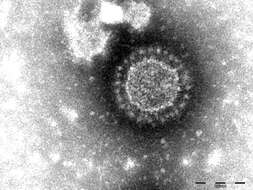
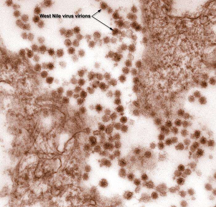
Description: English: This is a digitally-colorized transmission electron microscopic (TEM) image revealed the presence of West Nile virus (WNV) virions, in an isolate that was grown in a cell culture. Date: Unknown dateUnknown date. Source: https://phil.cdc.gov/Details.aspx?pid=10700. Author: Cynthia Goldsmith.
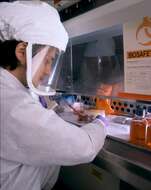
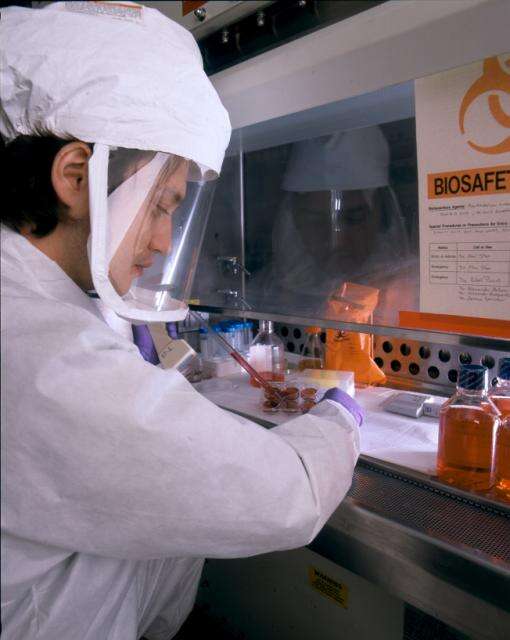
Description: NIAID scientist wears BSL-3 gear to demonstrate preparation of tissue culture plates infected with a chimera of West Nile virus and the dengue virus. The lab is developing a live, attenuated virus vaccine for West Nile virus. Credit: NIAID. Date: Taken on 27 April 2011, 12:50. Source: West Nile Vaccine Research in BSL-3 Lab. Author: NIAID. Camera location39° 00′ 03.27″ N, 77° 05′ 57.49″ WView all coordinates using: OpenStreetMap 39.000909; -77.099304.
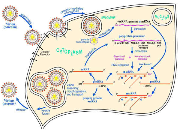
All images in this article were uploaded in the JPEG format even though it consists of non-photographic data. This information could be stored more efficiently or accurately in the PNG or SVG format. If possible, please upload a PNG or SVG version of this image without compression artifacts, derived from a non-JPEG source (or with existing artifacts removed). After doing so, please tag the JPEG version with {{Superseded|NewImage.ext}} and remove this tag. This tag should not be applied to photographs or scans. For more information, see {{BadJPEG}}. Description: English: West Nile virus life cycle. After binding and uptake, the virion envelope fuses with cellular membranes, followed by uncoating of the nucleocapsid and release of the RNA genome into the cytoplasm. The viral genome serves as messenger RNA (mRNA) for translation of all viral proteins and as template during RNA replication. Copies are subsequently packaged within new virus particles which are transported in vesicles to the cell membrane. Date: 26 May 2013, 21:37:46. Source: https://www.ncbi.nlm.nih.gov/pmc/articles/PMC3311072/. Author: De Filette, et al.
Description: English: Porcine epidemic diarrhea virus particles seen by negative-stain electron microscopy of fecal samples. Negative staining with 1% phosphotungstic acid. Scale bar indicates 100 nm. Date: 1 March 2015. Source: https://wwwnc.cdc.gov/eid/article/21/3/14-1165_article. Author: Dennis Hanke, Maria Jenckel, Anja Petrov, Mathias Ritzmann, Julia Stadler, Valerij Akimkin, Sandra Blome, Anne Pohlmann, Horst Schirrmeier, Martin Beer, and Dirk Höper.
Description: English: Porcine deltacoronavirus (OH-FD22) particle detected in intestinal contents from a gnotobiotic pig. The sample was negatively stained with 3% phosphotungstic acid. Scale bar indicates 100 nm. Date: 1 April 2015. Source: https://wwwnc.cdc.gov/eid/article/21/4/14-1859_article. Author: Kwonil Jung, Hui Hu, Bryan Eyerly, Zhongyan Lu, Juliet Chepngeno, and Linda J. Saif.
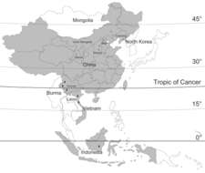
Description: English: Location of new Banna viruses (BAVs) isolated in China (red triangles) and previously reported BAV isolation sites (black triangles). Countries reporting isolation of BAV are shaded. The names of the countries that are contiguous with BAV isolation sites are labeled. BAV distribution sites in Indonesia, Vietnam, and part of China are located in tropical zones, which lie predominantly between the Tropic of Cancer and the equator. Most BAV distribution sites in China in the area from the Tropic of Cancer to latitude 45°N belong to the northern temperate zone. Date: 8 June 2013, 02:57:43. Source: http://wwwnc.cdc.gov/eid/article/16/3/09-1160_article.htm. Author: Liu, H., et al.
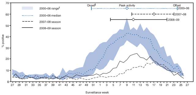
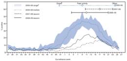
Description: English: Percentage of rotavirus tests with positive results, by surveillance week, United States, July 2000--June 2009. Date: 2009. Source: https://www.cdc.gov/mmwr/preview/mmwrhtml/mm5841a2.htm. Author: U.S. Department of Health and Human Services.
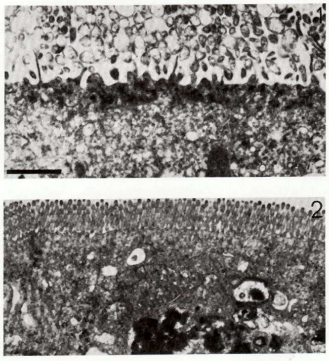
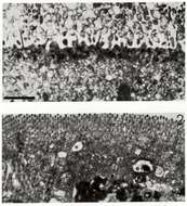
Description: Transmission electron micrograph of an enterocyte 3 days after infection by rotavirus (top). The microvilli have been destroyed. An unifected enterocyte is shown for comparison at the bottom. The bar = approx 500 nm. Date: 5 November 2007. Source: English Wikipedia. Author: Graham Beards. English Wikipedia user Graham Beards, the copyright holder of this work, hereby publishes it under the following license: : Permission is granted to copy, distribute and/or modify this document under the terms of the GNU Free Documentation License, Version 1.2 or any later version published by the Free Software Foundation; with no Invariant Sections, no Front-Cover Texts, and no Back-Cover Texts. A copy of the license is included in the section entitled GNU Free Documentation License.http://www.gnu.org/copyleft/fdl.htmlGFDLGNU Free Documentation Licensetruetrue. : This file is licensed under the Creative CommonsAttribution 3.0 Unported license.:. You are free: to share – to copy, distribute and transmit the work to remix – to adapt the work Under the following conditions: attribution – You must attribute the work in the manner specified by the author or licensor (but not in any way that suggests that they endorse you or your use of the work). http://creativecommons.org/licenses/by/3.0 CC BY 3.0 Creative Commons Attribution 3.0 truetrue.
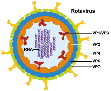
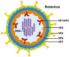
Summary.mw-parser-output table.commons-file-information-table,.mw-parser-output.fileinfotpl-type-information{border:1px solid #a2a9b1;background-color:#f8f9fa;padding:5px;font-size:95%;border-spacing:2px;box-sizing:border-box;margin:0;width:100%}.mw-parser-output table.commons-file-information-table>tbody>tr,.mw-parser-output.fileinfotpl-type-information>tbody>tr{vertical-align:top}.mw-parser-output table.commons-file-information-table>tbody>tr>td,.mw-parser-output table.commons-file-information-table>tbody>tr>th,.mw-parser-output.fileinfotpl-type-information>tbody>tr>td,.mw-parser-output.fileinfotpl-type-information>tbody>tr>th{padding:4px}.mw-parser-output.fileinfo-paramfield{background:#ccf;text-align:right;padding-right:0.4em;width:15%;font-weight:bold}.mw-parser-output.commons-file-information-table+table.commons-file-information-table,.mw-parser-output.commons-file-information-table+div.commons-file-information-table>table{border-top:0;padding-top:0;margin-top:-8px}@media only screen and (max-width:719px){.mw-parser-output table.commons-file-information-table,.mw-parser-output.commons-file-information-table.fileinfotpl-type-information{border-spacing:0;padding:0;word-break:break-word;width:100%!important}.mw-parser-output.commons-file-information-table>tbody,.mw-parser-output.fileinfotpl-type-information>tbody{display:block}.mw-parser-output.commons-file-information-table>tbody>tr>td,.mw-parser-output.commons-file-information-table>tbody>tr>th,.mw-parser-output.fileinfotpl-type-information>tbody>tr>td,.mw-parser-output.fileinfotpl-type-information>tbody>tr>th{padding:0.2em 0.4em;text-align:left;text-align:start}.mw-parser-output.commons-file-information-table>tbody>tr,.mw-parser-output.fileinfotpl-type-information>tbody>tr{display:flex;flex-direction:column}.mw-parser-output.commons-file-information-table+table.commons-file-information-table,.mw-parser-output.commons-file-information-table+div.commons-file-information-table>table{margin-top:-1px}.mw-parser-output.fileinfo-paramfield{box-sizing:border-box;flex:1 0 100%;width:100%}} Description: A rotavirus is a wheel-shaped virus that gets its name from its complex shape. Its genome consists of 11 double-stranded RNA segments that generate six structural proteins (VP1, VP2, VP3, VP4, VP6 & VP7) and six nonstructural proteins (NSP1-6). Each virus particle is surrounded by a triple layer coat. Credit: NIAID. Date: 21 October 2010, 13:52. Source: Rotavirus. Author: NIAID.
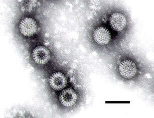
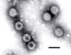
Description: English: Note the wheel-like appearance of some of the rotavirus particles. The observance of such particles gave the virus its name ('rota' being the Latin word meaning wheel). Bar = 100 nanometers. Source: Cell culture. Method: Negative-stain Transmission Electron Microscopy. Source: http://www.epa.gov/microbes/rota.html. Author: F.P. Williams, U.S. EPA.
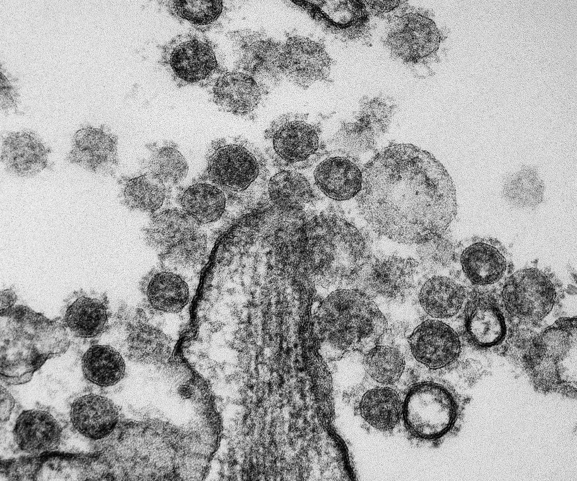
Summary.mw-parser-output table.commons-file-information-table,.mw-parser-output.fileinfotpl-type-information{border:1px solid #a2a9b1;background-color:#f8f9fa;padding:5px;font-size:95%;border-spacing:2px;box-sizing:border-box;margin:0;width:100%}.mw-parser-output table.commons-file-information-table>tbody>tr,.mw-parser-output.fileinfotpl-type-information>tbody>tr{vertical-align:top}.mw-parser-output table.commons-file-information-table>tbody>tr>td,.mw-parser-output table.commons-file-information-table>tbody>tr>th,.mw-parser-output.fileinfotpl-type-information>tbody>tr>td,.mw-parser-output.fileinfotpl-type-information>tbody>tr>th{padding:4px}.mw-parser-output.fileinfo-paramfield{background:#ccf;text-align:right;padding-right:0.4em;width:15%;font-weight:bold}.mw-parser-output.commons-file-information-table+table.commons-file-information-table,.mw-parser-output.commons-file-information-table+div.commons-file-information-table>table{border-top:0;padding-top:0;margin-top:-8px}@media only screen and (max-width:719px){.mw-parser-output table.commons-file-information-table,.mw-parser-output.commons-file-information-table.fileinfotpl-type-information{border-spacing:0;padding:0;word-break:break-word;width:100%!important}.mw-parser-output.commons-file-information-table>tbody,.mw-parser-output.fileinfotpl-type-information>tbody{display:block}.mw-parser-output.commons-file-information-table>tbody>tr>td,.mw-parser-output.commons-file-information-table>tbody>tr>th,.mw-parser-output.fileinfotpl-type-information>tbody>tr>td,.mw-parser-output.fileinfotpl-type-information>tbody>tr>th{padding:0.2em 0.4em;text-align:left;text-align:start}.mw-parser-output.commons-file-information-table>tbody>tr,.mw-parser-output.fileinfotpl-type-information>tbody>tr{display:flex;flex-direction:column}.mw-parser-output.commons-file-information-table+table.commons-file-information-table,.mw-parser-output.commons-file-information-table+div.commons-file-information-table>table{margin-top:-1px}.mw-parser-output.fileinfo-paramfield{box-sizing:border-box;flex:1 0 100%;width:100%}} Description: Transmission electron micrograph of Middle Eastern Respiratory Syndrome CoV particles found near the periphery of an infected MRC-5 cell. Credit: NIAID. Date: 19 July 2013, 12:39. Source: MERS Coronavirus Particles. Author: NIAID.
Sin-Yee Fung, Kit-San Yuen, Zi-Wei Ye, Chi-Ping Chan, and Dong-Yan Jin
Wikimedia Commons
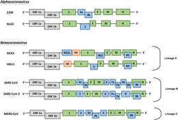
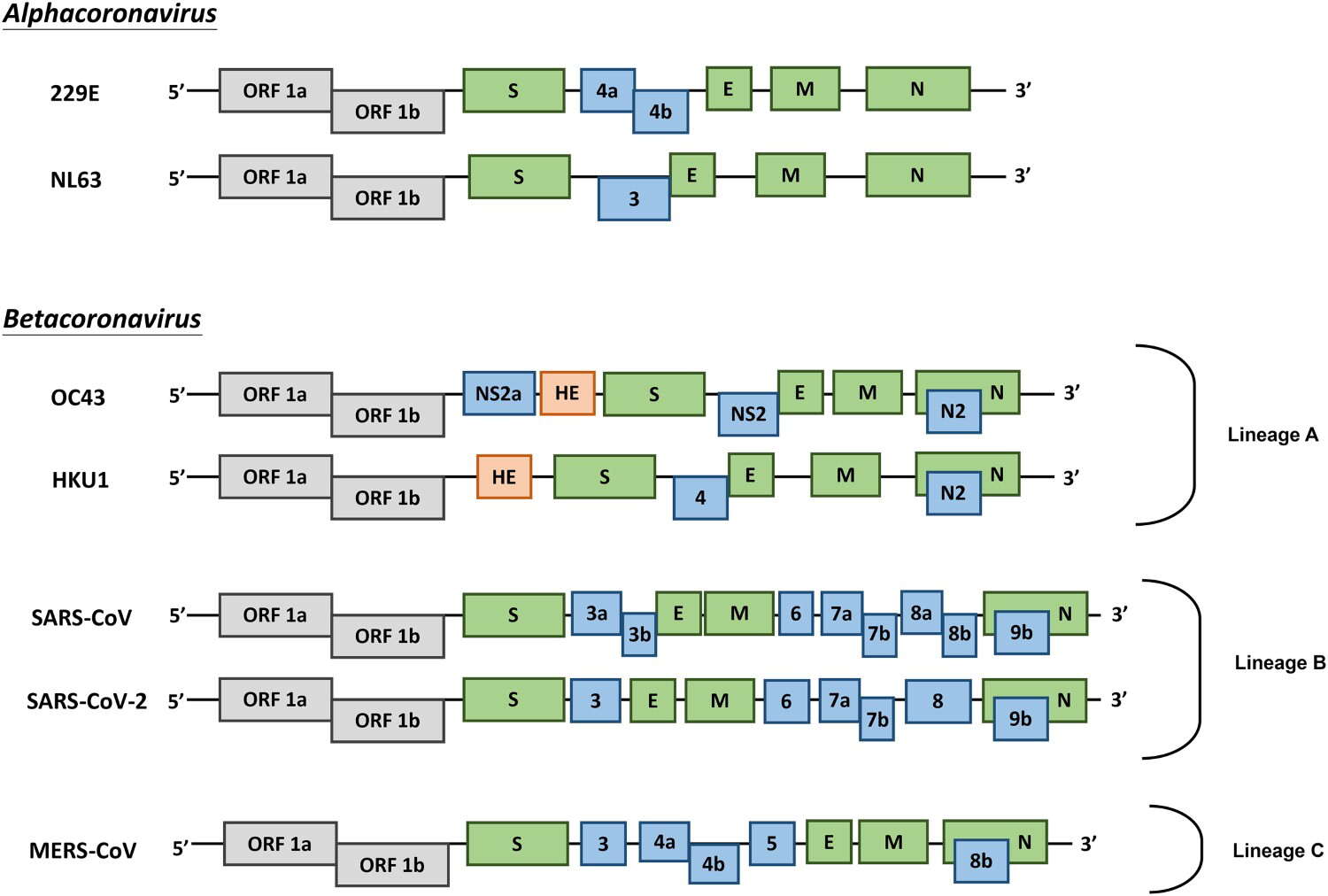
Description: English: Genome organization of HCoVs. Schematic diagram of seven known HCoVs is shown (not in scale). The genes encoding structural proteins spike (S), envelope (E), membrane (M), and nucleocapsid (N) are in green. The gene encoding haemagglutinin-esterase (HE) in lineage A of betacoronaviruses is in orange. The genes encoding accessory proteins are in blue. Date: 14 March 2020. Source: https://www.tandfonline.com/doi/full/10.1080/22221751.2020.1736644. Author: Sin-Yee Fung, Kit-San Yuen, Zi-Wei Ye, Chi-Ping Chan, and Dong-Yan Jin.
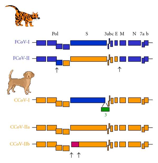
Description: English: Genetic relationships between the different feline and canine coronaviruses genotypes (FCoV and CCoV). The feline sequences are coloured in blue, the canine sequences in orange, and the porcine sequences in purple. Arrows indicate the putative sites of recombinations. The genes encoding for the polymerase polyprotein (pol), the structural spike (S), the envelope (E), the membrane (M), and the nucleocapsid (N) proteins are indicated. The genes encoding the accessory proteins are designated by numerals. Date: 31 July 2011. Source: https://www.hindawi.com/journals/av/2011/609465/. Author: Sophie Le Poder.


