-
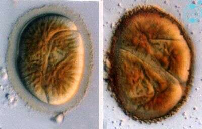
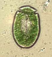
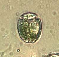
Amphidinium (am-fee-din-ee-um) britannicum (Herdman) Lebour 1925. The image on the left shows a cell in ventral view. The cingulum is strongly descending and therefore the epicone is asymmetrical. The chloroplasts are yellow-brown. These cells are not motile stage (temporary cysts) surrounded by a hyaline layer. The image on the right shows a dividing cell surrounded by granules.
-
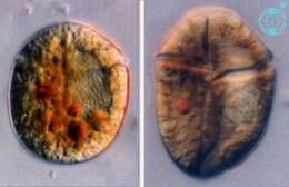
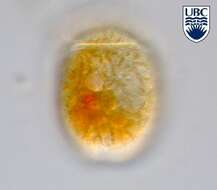
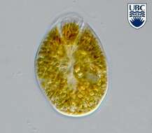
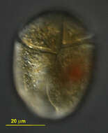
Amphidinium (am-fee-din-ee-um) corpulentum Kofoid & Swezy 1921. The image on the left is a mid focus plane through a cell showing the nucleus on the right side of the cell (= the right image side). Red to orange food vacuoles and yellow-brown plastids are visible. The image on the right shows a cell in ventral view (side-reversed). The cingulum is at the anterior end and an apical groove is visible.
-
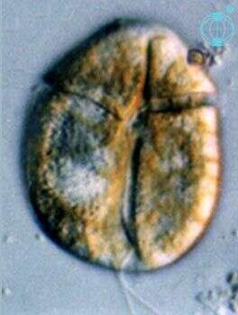
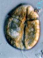
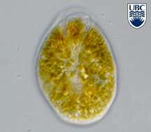
Amphidinium (am-fee-din-ee-um) corpulentum Kofoid & Swezy 1921. The image shows a cell in ventral view. The nucleus is on the right cell side (left image side). The cingulum is near the anterior end of the cell. The apical groove is clearly visible. The platids are yello-brown.
-
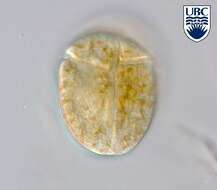
Amphidinium corpulentum Kofoid et Swezy 1921.
-
Amphidinium corpulentum Kofoid et Swezy 1921.
-
Amphidinium corpulentum Kofoid et Swezy 1921.
-
Amphidinium corpulentum Kofoid et Swezy 1921.
-
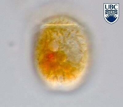
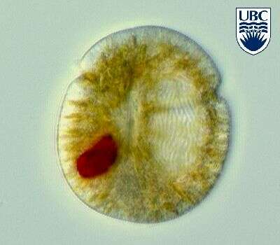
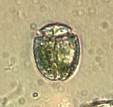
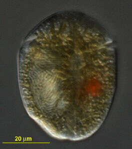
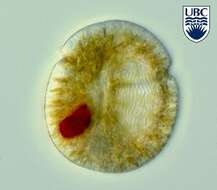
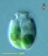
Amphidinium (am-fee-din-ee-um) latum Lebour 1925. The image shows a cell in ventral view. The red stigma is visible in the sulcal area. The plastids are blue-green and yellow-brown. The epicone is shorter than the hypocone.
-

Cells rounded square to oblong from the ventral side, dorso-ventrally flattened. Length 16 - 25 microns, width 13 - 26 microns, length to width ratio 0.8 - 1.8. During ingestion, cell shape distorted, becoming broader and rounder. Epicone cone shaped, with a pointed apex. Cingulum wide, approximately 2 microns, completely encircling the cell. Sulcus narrow, a straight line down the middle of the cell, becoming wider at the antapex, where it forms a notch. Short sulcal extension present on the epicone. Apical groove present, continuing from the sulcal extension to the left of the apex and then in an anticlockwise spiral around the apex. Nucleus in the centre or to the left of the hypocone, rounded. Chloroplasts not present. Different coloured food bodies/kleptochloroplasts present, including bright pale green, brown and blue-green cells. Generally very fast swimming. Non-motile cells rarely observed, round, approximately 22 microns diameter, surrounded by a hyaline layer, cingulum often still recognisable.
-
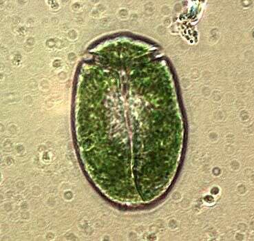
Amphidinium latum, an unsually large cell with green inclusions, observed in marine muds and sandy sediments in the vicinity of Broome, Western Australia in September 2003. This work was supported by the Australian Biological Resources Study.
-
Amphidinium latum observed in marine muds and sandy sediments in the vicinity of Broome, Western Australia in September 2003. This work was supported by the Australian Biological Resources Study.
-
Amphidinium latum, an unsually large cell with green inclusions, observed in marine muds and sandy sediments in the vicinity of Broome, Western Australia in September 2003. This work was supported by the Australian Biological Resources Study.
-
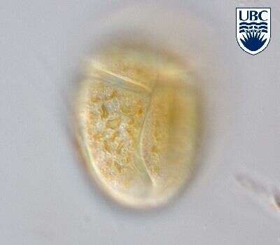
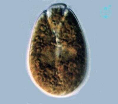
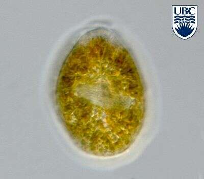
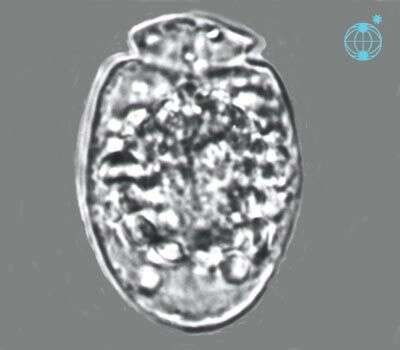
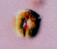
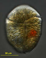
Amphidinium (am-fee-din-ee-um) mootonorum Murray et Patterson 2002. The image shows a cell in ventral view. The epicone is small. The plastids are yellow-brown. The nucleus is visible in the middle of the cell.
-
Cells oval from the ventral side, dorso-ventrally flattened. Length 30 - 50 microns, width 24 - 44 microns. Epicone flattened anteriorly, reaching to approximately 0.4 of the cell length from the apex, becoming narrow and 'stem shaped' at the junction of the cingulum and sulcus. Epicone slightly deflected towards the left, just visible above the hypocone on the dorsal side. Cingulum relatively wide (2 - 3 microns), proximal end 0.4 of the cell length from the apex, travelling up to 0.05 of the cell length from the apex on the dorsal side, then descending, ends not displaced. Sulcus narrow initially, opening into a tear-drop shaped indentation approximately 0.7 of the cell length from the apex, not reaching the antapex. Short (2 - 3 microns) groove extends vertically near the ventral posterior end of the epicone . Nucleus in the centre of the hypocone, elongate oval. Non-motile cells slightly broader than motile cells, surrounded by hyaline layer, hypocone completely surrounds the epicone at the apex of the cell. Many small (2 - 4 microns diameter), yellow-brown chloroplasts present. Small lipid globules often present. Very occasionally, red bodies, possibly food particles, present. In high focus, surface of cell appears rough or grainy. A clonal culture of this species was temporarily kept. Under the culture conditions used, non-motile cells numerically dominated the culture over motile cells. Dividing non-motile cells were also seen.
-
Amphidinium mootonorum Murray et Patterson 2002.
-
Amphidinium mootonorum Murray et Patterson 2002.
-
Amphidinium mootonorum Murray et Patterson 2002.
-
Cells oval from the ventral side, dorso-ventrally flattened. Length 20 - 28 microns, width 10 - 20 microns, length to width ratio 1.3 - 2.0. Epicone triangular, curved anteriorly, deflected to the left. Cingulum beginning 0.2 of the cell length from the apex, distal end 2 - 4 microns below and to the right of the proximal. Sulcus beginning just to the right of the mid-ventral line, initially deep and wide (2 - 3 microns), becoming less distinct as it nears the posterior of the cell. Two pusules present, each approximately 1 microns diameter, one below the origin of the cingulum, the to the right of the origin of the sulcus. Longitudinal flagellum arising in a pocket just to the left of and below the origin of the sulcus. Nucleus in the posterior part of the hypocone, round to oval, approximately 10 microns diameter. Plastids yellow-brown, in strands radiating from the central pyrenoid, 4 -5 microns diameter . Asexual division occurring in hyaline covered cysts, in which either two or three daughter cells may be formed.
-
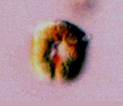
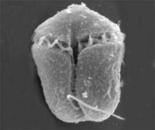
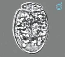
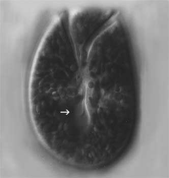
A gymnodinioid dinoflagelate, identified by Shauna Murray, isolated from sandy sediments and photographed by Bob Moore and Dan Lahr. The nucleus with very dense genetic material is very evident in his picture.
-
A optical slice showing some of the grooves in this gymnodinioid dino. Identifyed by Shauna Murray, Isolated and photographed by Bob Moore and Dan Lahr.
-
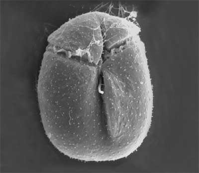
Evident grooves on this vicious predator. Identified by Dr. Murray, isolated by Bob Moore, picture by Dan Lahr.
-
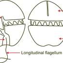
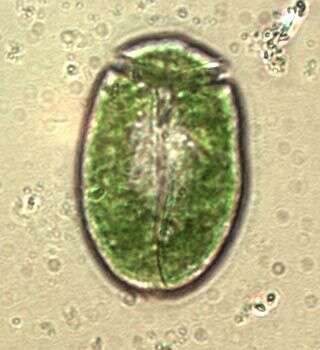
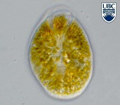
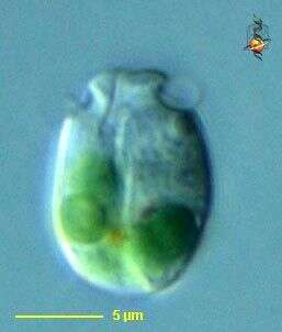
This species (A. poecilochroum?) contains a number of symbiotic blue green algae (identifiable as such from the colour). Many protists contain blue green algae, and this has been used by some to create a taxon for protists with these symbionts, imagining that they represent some kind of intermediary between bacterial and eukaryotic life. The equatorial flagellum can be seen as it curves round the cell, the groove in which the longitudinal flagellum normally resides is evident, although the flagellum is not visible.
-
Amphidinium (am-fee-din-ee-um) poecilochroum Larsen 1985. The image shows a cell in ventral view. The epicone is small, the cingulum is near the anterior end of the cell.
-
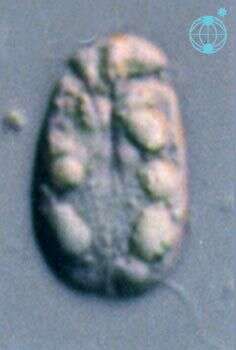
Amphidinium (am-fee-din-ee-um) poecilochroum Larsen 1985. The image shows a mid-focal plane through a cell. The epicone is small, the cingulum is near the anterior end of the cell.