nimet breadcrumb-navigoinnissa
“Drepanodontus tatyanae new species
(Figures 24-50, Table 2)
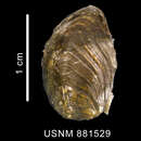
Description: Shell (Figures 24-30, 33-35) large (to 43.1 mm), thin, strongly fusiform. Protoconch eroded on all specimens. Early whorls of holotype (Figure 27) indicate that protoconch would likely have been ~2-2 ½ whorls, < 3 mm in diameter. Transition to teleoconch indistinct in holotype. Teleoconch estimated to consist of up to 7 whorls. Early teleoconch whorls slightly concave in profile, with narrow spiral cords. By third whorl, teleoconch becomes convex, increasingly so in subsequent whorls, forming evenly rounded, oval whorls without distinct shoulder. Suture abutting onto pronounced peripheral cord (Figures 24, 33, arrow). Axial sculpture of pronounced growth lines, opisthocyrt on early whorls, becoming weaker, orthocline by fourth teleoconch whorl. Spiral sculpture of broad, sharp cords (9-12 on penultimate whorl, 10-14 from suture to peripheral cord, 16-24 from peripheral cord to tip of siphon) narrower than intervening spaces, with 0-4 very fine threads between adjacent cords, especially near periphery. Peripheral cord, slightly thicker more pronounced that others, demarcates inflection in curvature of outer lip, evenly convex abapically, evenly concave from peripheral cord to tip of siphonal canal. Aperture large (AL/SL = 0.56), roughly elliptical, deflected from shell axis by 13-17°. Siphonal canal long (~1/4 shell length), broad, open, crosses shell axis. Outer lip thin, not reflected. Inner lip of weakly concave parietal region, slightly longer columella, long siphonal fold extending beyond glossy, translucent parietal region, broad posteriorly, tapering toward base of siphonal canal. Neither siphonal fasciole nor pseudoumbilicus present. Shell uniformly white, aperture, parietal callus glossy. Periostracum straw-colored, of widely spaced, short axial lamellae. Operculum (Figures 31-32, 41-42, op), large (~0.82 AL), oval, thin, brownish yellow, flexible, with terminal nucleus. Attachment of columellar muscle clearly visible through operculum, oval, spans about 2/3 of operculum surface.
Anatomy (Paratype 1): Soft tissues (Figures 41-42) comprise approximately 3 ½ whorls. Mantle cavity spans slightly less than ½ whorl. Kidney (Figures 41, 42, nep) broad, spans slightly less than ½ whorl, with 12 transverse folds of unequal width visible through wall. Nephridial gland (Figure 41, ng) narrow, situated anterodorsally to the nephridium. Digestive glands (Figures 41, 42, adg, pdg) of 2 ½ whorls. Columellar muscle (Figure 42, cm) of 1 ½ whorls, attached to shell at rear of mantle cavity. Foot medium-sized, short in contracted specimen (L/W ~1.2), with well-developed narrow propodium (Figure 42, prp) separated by narrow propodial cleft. Body color yellowish tan, without pigmentation. Head medium-sized, with short, stout, conical tentacles, with small but distinct lobes at their bases. Eyes present (Figure 42, e), light gray in color and semitransparent, deeply embedded into lobes. Mantle does not cover base of head.
Mantle Cavity (Figure 48): Mantle cavity deep (L/ W ~1.2). Mantle edge smooth, slightly thickened. Siphon (Figure 48, s) short, free, muscular, broad, extending well beyond mantle edge, with thick siphonal edge (Figure 48, se), covering anteriormost part of ctenidium. Osphradium (Figure 48, os) greenish, bipectinate, large, wide, ½ as long, 2/3 as wide as ctenidium. Ctenidium (Figure 48, ct) large, curved, spanning nearly entire mantle cavity length. Ctenidial lamellae broad, triangular, with short recurved edges along posterior part (closer to pericardium), gradually becoming narrower, relatively taller anteriorly. Hypobranchial gland lacks distinct folds, covered by thick layer of mucus. Rectum long, spanning ~4/5 of mantle cavity length.
Table 2. Drepanodontus tatyanae new species. Measurements of shell characters. Linear measurements in mm. (n = 3, including holotype).
Character
Mean
σ
Range
Holotype
Shell length (SL)
45.1
10.1
36.2-56.0
43.1
Final whorl length (FWL)
33.8
7.4
28.6-42.2
30.5
Aperture length (AL)
26.7
6.2
22.2-33.7
24.1
Siphonal canal length (SCL)
11.4
4.7
7.5-16.7
10.1
Shell width (SW)
19.1
2.3
17.5-21.8
18.1
FWL/SL
0.75
0.04
0.71-0.79
0.71
AL/SL
0.59
0.03
0.56-0.61
0.56
SCL/SL
0.25
0.05
0.21-0.30
0.23
SW/SL
0.43
0.05
0.39-0.48
0.42
Number of spiral cords on penultimate whorl
10.0
1.5
9-12
10
Number of spiral cords suture to peripheral cord
12.0
2.0
10-14
12
Number of spiral cords peripheral cord to siphon
19.7
4.0
16-24
19
Alimentary System (Figures 41-47): Proboscis non-pigmented, (Figures 43, 45, pr) short when retracted (~0.2 SL, 0.58 AL), thick (L/D ~3), with slightly folded walls. Proboscis retractor muscles (Figures 45, 46, prr) not numerous, thin, attached to thin-walled, translucent proboscis sheath at middle-posterior region when proboscis retracted. Proboscis wall thin, ~1/10 proboscis diameter. Anterior part of proboscis flattened to form rim surrounding mouth opening (Figure 43, mo) in form of irregular triangular slit. Anterior oesophagus very broad, nearly filling proboscis. Dorsal folds very large, bordering deep groove. Buccal mass small, spans slightly more than ½ of proboscis length. Odontophoral cartilages paired, fused anteriorly, extend nearly entire length of buccal mass, but < ½ proboscis length. Radular ribbon (Figures 36-40) equal in length to cartilages, 6.6 mm (0.35 AL), about 380 µm wide (0.020 AL), triserial, consisting of 50 rows of teeth, posteriormost 5 rows nascent. Rachidian teeth of Paratype 1 (Figures 36-37) with 4 cusps, here interpreted as comprising a long central cusp, flanked by shorter, outer cusps, with an additional, asymmetrical cusp on the left side. Rachidian teeth of a second specimen (Figures 38-40) appear monocuspid, but "central cusp" consists of 3 incompletely fused cusps, flanked by additional small denticles (Figure 40). Lateral teeth with distinctive shape, with single large, recurved outer cusp emanating from long, narrow basal plate. Inner surface of cusp with 1 or more, occasionally bifid, posteriorly directed denticles, with number, size of denticles varying from side to side and along radular ribbon.
Right salivary gland medium-sized, rounded, partially covering valve of Leiblein (Figures 43, 44, rsg), laterodorsal to nerve ring. Left salivary gland slightly smaller than right, irregularly shaped, dorsal to nerve ring, ventral to proboscis with its main axis perpendicular to proboscis axis, appears small when viewed from left (Figure 45, lsg). Salivary ducts (Figures 44, 46, sd) short, thick, enter oesophagus wall shortly after leaving gland. Valve of Leiblein (Figure 46, vL) well defined, large, pyriform, with whitish glandular pad visible through walls of valve.
Gland of Leiblein (Figures 43-45, gL) yellowish, slightly darker than other organs of cephalic haemocoel, medium sized, long, tubular, coiled anteriorly. Gland thin-walled, ascinous anteriorly (Figure 46, agL), opens into oesophagus slightly posterior to nerve ring via broad, short duct (Figure 46, dgL), becomes thinner, more transparent posteriorly (Figure 46, pgL), tapering to become flaccid, non-glandular (Figure 46, vgL).
Oesophagus thick, broad anterior to nerve ring, narrowing slightly posterior to the ring. Posterior oesophagus expands greatly to form "crop," (Figure 47, poe) then gradually narrows towards opening into stomach. Stomach (Figure 47) very large, spans ~ ½ whorl, from the posterior border of nephridium, U-shaped, without posterior mixing area. Preservation inadequate to discern internal morphology. Digestive glands ducts (Figure 47, ddg) large, paired, closely spaced. Posterior duct close to oesophagus entrance, anterior duct at mid-length of stomach. Digestive glands clearly separate. Anterior gland small (Figure 41, adg), spans - ½ whorl, posterior gland ~2 whorls (Figures 41, 42, pdg). Glands meet at the level of the posterior duct to digestive gland. Rectum long, spans ~4/5 of mantle cavity length. Rectum thin-walled, very broad, filled with polychaete spicules, numerous sand grains of different sizes.
Female Reproductive System: Paratype 1, mature female. Pallial gonoduct consists of long, tubular, capsule gland (Figures 42, 48, cg), with a small bursa copulatrix anterior to it. Genital opening (Figure 48, go) below, slightly posterior to anus (Figure 48, a).
Male Reproductive System: Paratype 2, male. Penis (Figure 49) long, very narrow, flattened laterally. Seminal papilla very small, blunt, surrounded by deep circular fold around its base.
Type Locality: NE of South Shetland Islands, 59°01' S, 52°00' W, in 3010-3510 m. [R/V ELTANIN cruise 22, Sta. 1511, 26 Jan 1966].
Type Material: Holotype, USNM 1010544, from the type locality. Paratype 1♀ , Paratype 2 ♂, USNM 881529, E of South Sandwich Islands, 57°00.24' S, 26°10.06' W, in 2740-2757 m. [R/V ELTANIN cruise 575, Sta. 38, 22 May 1975]
Other Material Examined: USNM 1010545, South Atlantic Ocean [Argentine Abyssal Plain], 47°17.3' S, 47°45.7' W, in 5685-5798 M. [R/V ISLAS ORCADAS Cruise 575, Sta. 4, 8 May 1975], 2 bodies without shells (radula illustrated, Figures 38-40); USNM 1010546, Scotia Sea, S of South Georgia Island, 58°04' S, 37°50' W, 3255-3166 m. [R/V ELTANIN cruise 9, sta. 699, 30 Aug 1963], 1 dead poorly preserved juvenile. USNM 1013084, Scotia Sea, SW of South Georgia Island, 55°56' S, 44°56' W, 3742-3614 m. [R/V ELTANIN cruise 575, Sta. 472, 13 Feb 1963], 1 body and fragments of the shell.
Distribution (Figure 50): This species occurs in the Scotia Sea and adjacent Argentine Abyssal Plain, at depths of 2740-5798 m.
Etymology: This species is named in honor of the junior author's wife, Tatyana Steiker, an ichthyologist and illustrator at the P. P. Shirsov Institute of Oceanology.
Remarks: The large, elongate, fusiform, siphonate, spirally corded shell of Drepanodontus tatyanae easily distinguishes this species from most Antarctic buccinoideans. Conchological similarity is limited to relatively few large taxa, notably Antarctoneptunea aurora (Hedley, 1912) and Cavineptunea monstrosa Powell, 1951, both members of continental shelf and upper slope faunas. Drepanodontus tatyanae is most easily distinguished from the former by having a distinctive peripheral spiral cord that demarcates a change in the direction of curvature in the outer lip, while the monotypic Cavineptunea is most easily distinguished by its unique, cylindrical, flat-sided, indented protoconch. Both Antarctoneptunea (Dell, 1972: fig. 6) and Cavineptunea (Powell, 1951: 145) have radulae with tricuspid rachidian teeth and lateral teeth with 3 (or 4) cusps, quite unlike the distinctive radula of Drepanodontus.
The radula of Drepanodontus tatyanae most closely resembles that of Kapala bathybius Bouchet and Warén, 1986 (Bouchet and Warén, 1986: fig. 8), a species inhabiting the Cape Basin off southwestern Africa at depths of 3550 m. Like Drepanodontus, K. bathybius has rachidian teeth that may appear to be monocuspid in some individuals, with anteriorly indented, squarish basal plates, and lateral teeth characterized by a single, large, sickle-like cusp with secondary denticles that vary in number and prominence from side to side and from tooth to tooth. The shell of K. bathybius, and the related K. bonaespei (Barnard, 1963), also from the Cape Basin in 2504-3103 m, are comparable in size, and also elongate, fusiform and spirally corded, but broader (Bouchet and Warén, 1986: figs. 42, 43), and lack the distinctive peripheral cord of Drepanodontus. Barnard (1963: 432, fig. 6b) illustrates and describes the radula of K. bonaespei (which he described as a Neptunea) to have rectangular rachidian teeth with a "median cusp, sometimes a minute denticle on one side or on both sides" and lateral teeth "unequally bicuspid, with 2-5 tiny denticles between the two cusps, the denticles not always symmetrical."
Barnard (1963) assigned this species to the genus Neptunea Roding, 1798, because of the similarity of its lateral teeth with those of boreal buccinoideans, despite striking differences in the morphology of the shell and rachidian teeth. Bouchet and Warén, (1986: 464) also commented on lateral tooth similarities of Kapala (including its type species, the southern Australian bathyal species K. kengrahami Ponder, 1982) with the type species of the boreal genera Volutopsius Morch, 1857, Neoberingius Habe and Ito, 1965, Ancistrolepis Dall, 1895, and certain representatives of Japelion Dall, 1918. These boreal taxa have elongated, spatulate lateral teeth with a large outer cusp, a significantly smaller inner cusp, and a variable number of smaller denticles or cusps between them. We interpret the lateral teeth of Drepanodontus to be different, in that they have a single, large outer cusp, but lack the shorter inner cusp of the boreal taxa. While some of the denticles that frequently emerge from the inner edge of the cusp of Drepanodontus may be large enough to be confused with an inner cusp, the lateral teeth of the boreal species are fundamentally bicuspid, while those of Drepanodontus are fundamentally monocuspid. Interestingly, the radula of K. bathybius illustrated by Bouchet and Warén (1986: fig. 8) has monocuspid lateral teeth lacking denticles distally, but developing denticles proximally along the left side of the radula, while the lateral teeth on the right side have 1 large and 1-3 smaller denticles along the inner edge of the single cusp.
The operculum of Drepanodontus is large, ovate, and has a terminal nucleus. While all species of Kapala share this opercular morphology, it is not distinctive, but widespread throughout Buccinoidea.
Although the presence of large eyes is not surprising in the bathyal type species of Kapala (Ponder, 1982: fig. 2), their occurrence in the abyssal taxa K. bonaespei (Barnard, 1963: 432) and Drepanodontus tatyanae is noteworthy. Other anatomical features that are congruent between Drepanodontus tatyanae and Kapala kengrahami include a small buccal mass and odontophore, a large kidney, a large, well-developed valve of Leiblein, crop, and a simple stomach.
Drepanodontus tatyanae co-occurs with Muffinbuccinum catherinae at the type locality of that species.”
(Harasewych & Kantor, 2004: 7-13)
Drepanodontus tatyanae is a species of sea snail, a marine gastropod mollusk in the family Buccinidae.[2]
The species is demersal, living close to the sea bed. They are broadcast spawners. Drepandodontus tatyanae grow from embryos to trochophores to veligers before maturing into adults.[3]
This species is distributed in the Scotia Sea, South Atlantic.
Drepanodontus tatyanae is a species of sea snail, a marine gastropod mollusk in the family Buccinidae.
Drepanodontus tatyanae is een slakkensoort uit de familie van de Buccinidae.[1] De wetenschappelijke naam van de soort is voor het eerst geldig gepubliceerd in 2004 door Harasewych & Kantor.
Bronnen, noten en/of referentiesDrepanodontus tatyanae là một loài ốc biển, là động vật thân mềm chân bụng sống ở biển trong họ Buccinidae.[2]
Chúng phân bố ở Biển Scotia, Nam Đại Tây Dương.
Drepanodontus tatyanae là một loài ốc biển, là động vật thân mềm chân bụng sống ở biển trong họ Buccinidae.