-
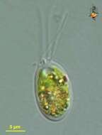
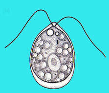
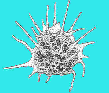
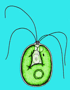
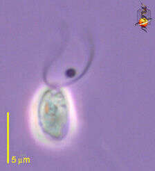
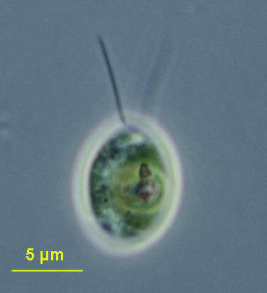
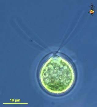
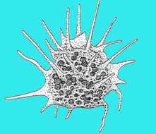
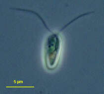
Portrait of Lobomonas stellata (Chodat), a volvocid flagellate. The ellipsoid to pear-shaped protoplast is separated from the cell wall by a space containing gelatinous material. The cell wall has irregularly spaced conical protrusions. There is one large cup-shaped chloroplast. A pyrenoid is located posteriorly. A peripheral stigma is located in the anterior 1/3 of the cell. Two equal flagella are about the length of the cell body. From freshwater pond near Boise, Idaho. Phase contrast.
-
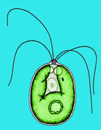
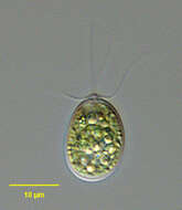
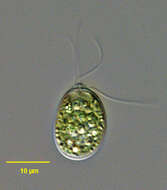
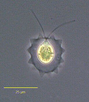
Portrait of Lobomonas stellata (Chodat), a volvocid flagellate. The ellipsoid to pear-shaped protoplast is separated from the cell wall by a space containing gelatinous material. The cell wall has irregularly spaced conical protrusions. There is one large cup-shaped chloroplast. A pyrenoid is located posteriorly. A peripheral stigma is located in the anterior 1/3 of the cell. Two equal flagella are about the length of the cell body. From freshwater pond near Boise, Idaho.DIC.
-
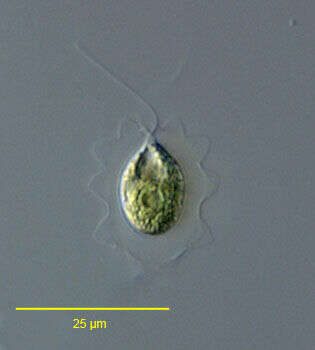
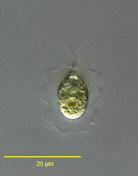
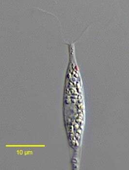
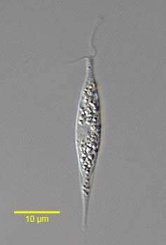
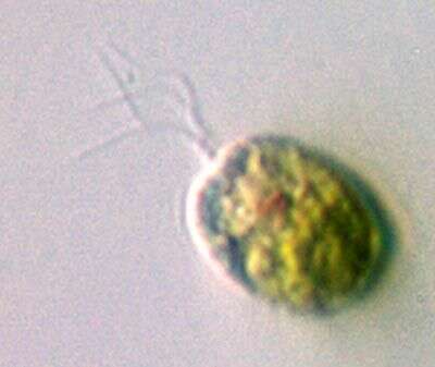
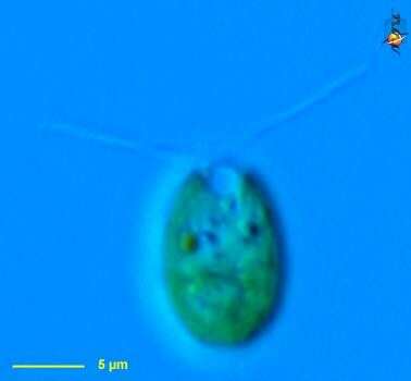
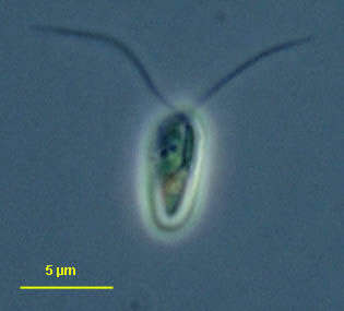
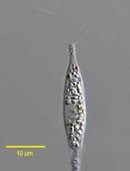
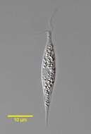
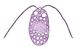
Hyalogonium klebsii (Klebs) Pascher,1927, a colorless volvocid flagellate with two equal anterior flagella. The body is elongate and fusiform. A red stigma is present on the left. Many small starch grains are seen in the cytoplasm.The nucleus is central. This genus may be confused with the euglenid Cyclidiopsis but Hyalogonium is smaller, biflagellate with thinner flagella, and lacks large paramylon bodies and the anterior canal opening. From standing rainwater pool near Boise, Idaho December 2005. DIC.
-
Hyalogonium klebsii (Klebs) Pascher,1927, a colorless volvocid flagellate with two equal anterior flagella. The body is elongate and fusiform. A red stigma is present on the left. Many small starch grains are seen in the cytoplasm.The nucleus is central. There are two anterior contractile vacuoles.This genus may be confused with the euglenid Cyclidiopsis but Hyalogonium is smaller, biflagellate with thinner flagella, and lacks large paramylon bodies and the anterior canal opening. From standing rainwater pool near Boise, Idaho December 2005. DIC.
-
-
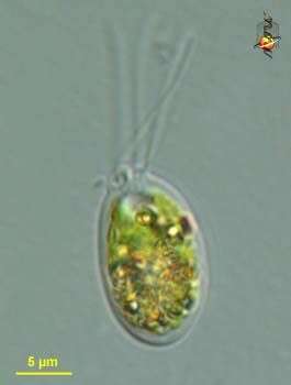
Carteria (car-tear-ee-a) a volvocid green algal cell, with chlorophyll b in the single cup-shaped plastid - giving it a green colour. Eyespot located in plastid, visible anterior of mid line on the right margin of the plastid. Very similar to Chlamydomonas, but distinguished by having 4 flagella. Differential interference contrast.
-
-
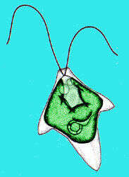
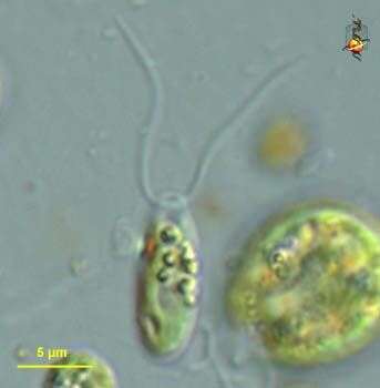
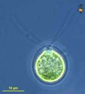
Portrait of the volvocid flagellate, Carteria (Diesing,1866). Two of the four equal length apical flagella are clearly seen inserting at the small anterior apical papilla. the other two are slightly out of the focal plane. The single large green plastid is cup-shaped. The small red eyespot is not seen well here.Collected from a freshwater pond near Boise, Idaho. DIC.
-
Portrait of the volvocid flagellate, Carteria (Diesing,1866). Two of the four equal length apical flagella are clearly seen inserting at the small anterior apical papilla. the other two are slightly out of the focal plane. The single large green plastid is cup-shaped.The two anterior contractile vacuoles are seen side by side just posterior to the apical papilla. The small red eyespot is not seen well here.Collected from a freshwater pond near Boise, Idaho. DIC.
-
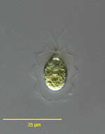
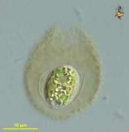
Pseudocarteria, a volvocid flagellate distinguished from the similar genus Carteria by absence of an anterior papilla. Four approximately equal-length flagella and single large chloroplast. Prominent stigma. From freshwater pond near Boise, Idaho. Oblique illumination.
-
-
-
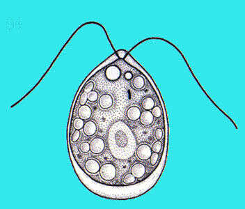
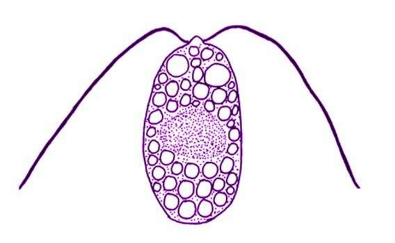
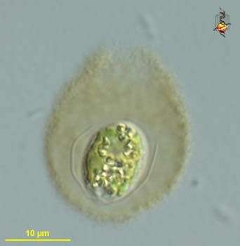
Polytoma papillata Pascher, 1927. Cells are 8-15 x 18-25 microns, cyst diameter20-30 microns Cell oval-ovoid with a conspicuous, hemispherical papilla, from which emerges two equal, isodynamic flagella. Two anterior contractile vacuoles, and a central nucleus. Many starch grains are scattered throughout the cell. The cytoplasm occasionally contains many differently coloured grains. No stigma. The swimming is slow and spiraling, and the cell may settle on the glass slide by the tip of the papilla. The species produces sporangia with 4 division products.
-
Chlamydomonas (clam-ee-doe-moan-ass), a solitary volvocid (flagellated green algal cell). Cell surrounded by a cellulosic wall, with two similar flagella emerging from near the apex. The photosynthetic pigments are located within a cup-shaped chloroplast which has a large pyrenoid with associated polysaccharide materials located posteriorly. The nucleus is located within the cup. Animations by Rosemary Arbur of flagellar beat patterns are available
here.Differential interference contrast.
-
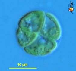
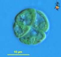
Chlamydomonas (clam-ee-doe-moan-ass), a solitary volvocid (flagellated green algal cell). Cell surrounded by a cellulosic wall, this is a division form in which four daughter cells are being produced at the same time. Animations by Rosemary Arbur of flagellar beat patterns are available
here. Differential interference contrast.
-
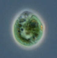
Chlamydomonas (clam-ee-doe-moan-ass), a solitary volvocid (flagellated green algal cell). Cell surrounded by a cellulosic wall, with two similar flagella emerging from near the apex. The photosynthetic pigments are located within a cup-shaped chloroplast which has a large pyrenoid with associated polysaccharide materials located posteriorly. The nucleus is located within the cup. This image shows the small red eyespot (orange colour here) and one anterior contractile vacuole. Differential interference contrast.
-
Chlamydomonas (clam-ee-doe-moan-ass), a solitary volvocid (flagellated green algal cell). Cell surrounded by a cellulosic wall, with two similar flagella emerging from near the apex. The photosynthetic pigments are located within a cup-shpaed chloroplast which has a large pyrenoid with associated polysaccharide materials. Many taxa described (there are books on this genus). Eyespot located within plastid. Flagella beat with a breast-stroke pattern. Phase contrast.
-
Chlamydomonas (clam-ee-doe-moan-ass), a solitary volvocid (flagellated green algal cell). Cell surrounded by a cellulosic wall, with two similar flagella emerging from near the apex. The photosynthetic pigments are located within a cup-shaped chloroplast which has a large pyrenoid with associated polysaccharide materials located posteriorly. The nucleus is located within the cup. This image shows one anterior contractile vacuole. Animations by Rosemary Arbur of flagellar beat patterns are available
here.Phase contrast.
-
Chlamydomonas (clam-ee-doe-moan-ass), a solitary volvocid (flagellated green algal cell). Cell surrounded by a cellulosic wall. Cell damaged, no flagella. The photosynthetic pigments are located within a cup-shaped chloroplast which has a large pyrenoid with associated polysaccharide materials located posteriorly. The nucleus is located within the cup. This image shows the red eyespot to the right and two anterior contractile vacuoles. Phase contrast.
-
Chlamydomonas (clam-ee-doe-moan-ass), a solitary volvocid (flagellated green algal cell). Cell surrounded by a cellulosic wall, with two similar flagella emerging from near the apex. Elongate species. Animations by Rosemary Arbur of flagellar beat patterns are available
here. Phase contrast.
-
Chlamydomonas (clam-ee-doe-moan-ass), a solitary volvocid (flagellated green algal cell). Cell surrounded by a cellulosic wall. With plastid containing chlorophyll B giving the bright green colour, two similar flagella emerge from the anterior of the cell. Differential interference contrast.
-
Chlamydomonas (clam-ee-doe-moan-ass), a solitary volvocid (flagellated green algal cell). Cell surrounded by a cellulosic wall and enclosed in mucilagenous sheath - cells in this form were inactive as if encysted. Differential interference contrast.
-
Chlamydomonas (clam-ee-doe-moan-ass), a solitary volvocid (flagellated green algal cell). Cell surrounded by a cellulosic wall. With plastid containing chlorophyll B giving the bright green colour, two similar flagella emerge from the anterior of the cell. Phase contrast.