-
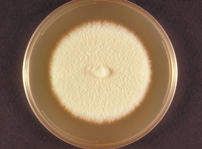
This is the top view of a Sabourauds dextrose plate culture growing the fungus Microsporum persicolor.Created: 1973
-
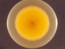
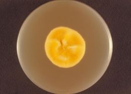
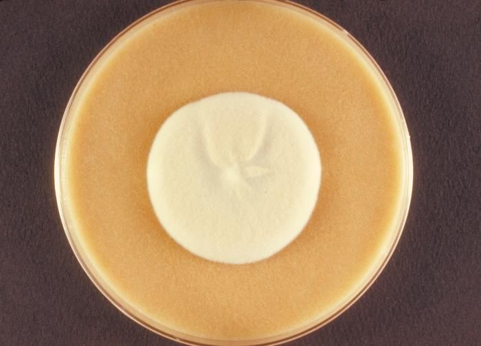
This is the bottom or reverse view of a cereal agar plate culture growing the fungus Microsporum persicolor.Created: 1973
-
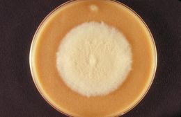
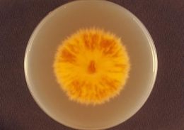
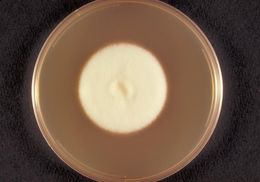
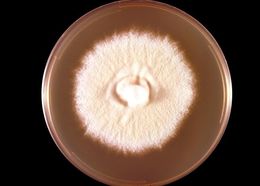
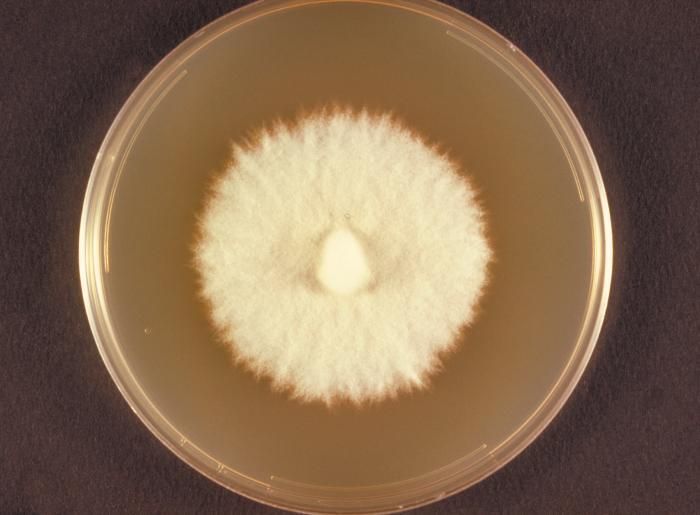
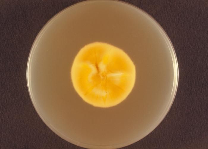
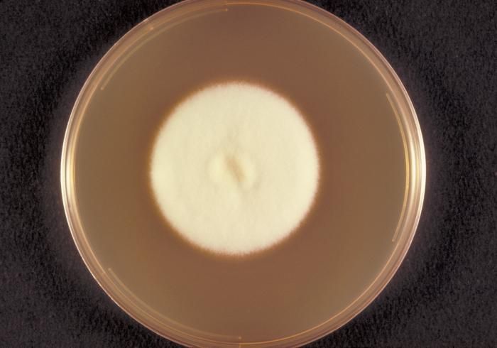
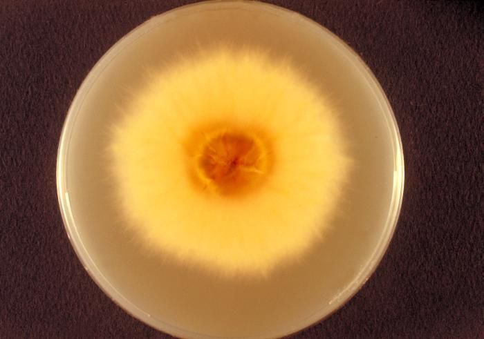
This is a top view of a cereal agar plate culture growing the fungus Microsporum persicolor.Created: 1973
-
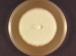
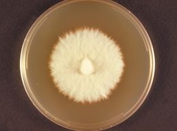
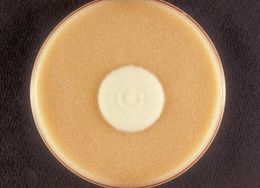
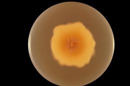
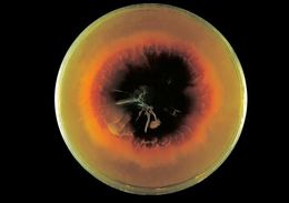
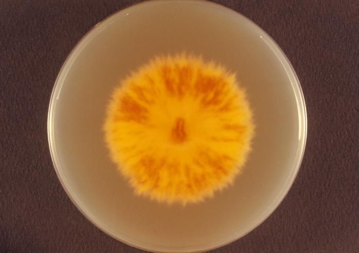
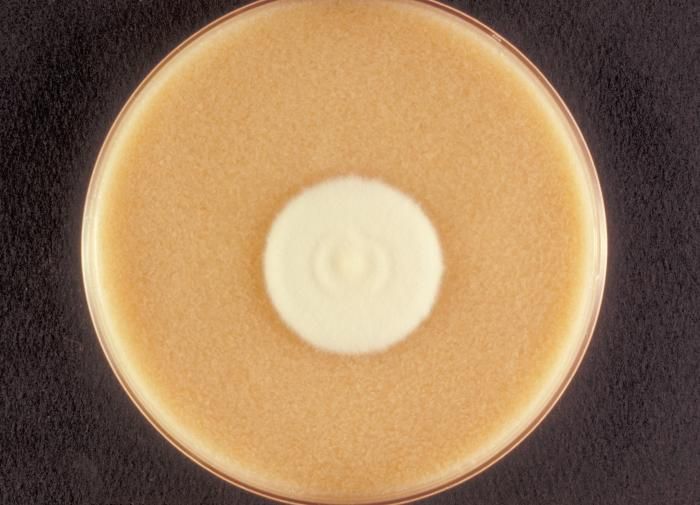
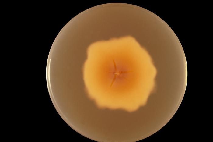
This is a top view of a Sabourauds dextrose agar plate culture growing Microsporum persicolor.Created: 1973
-
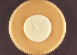
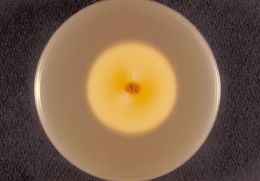
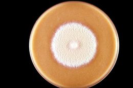
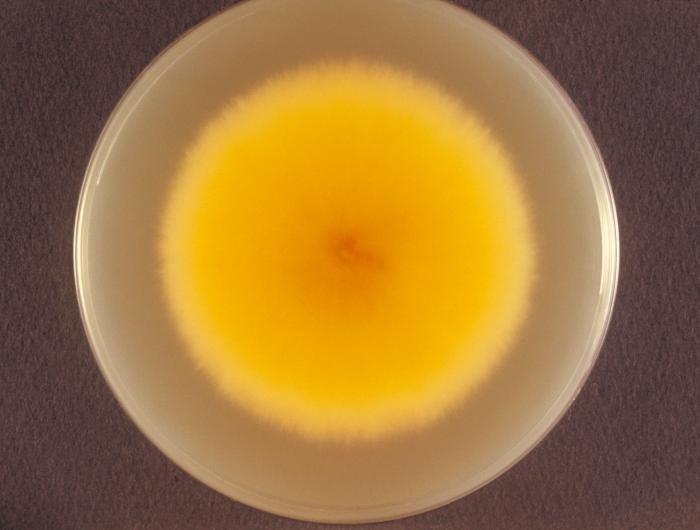
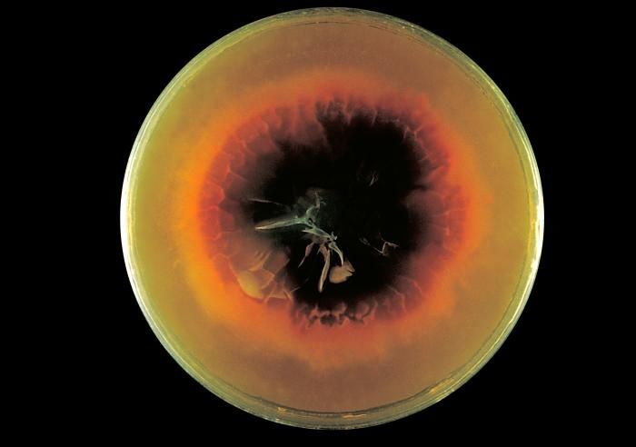
This is the bottom or reverse view of a Sabourauds dextrose plate culture growing the fungus Microsporum persicolor.Created: 1973
-
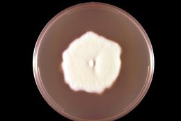
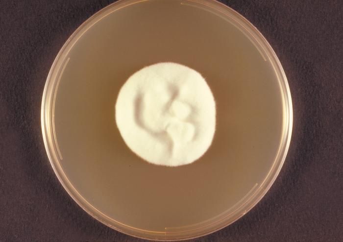
This is a top view of a cereal agar plate culture of a Microsporum persicolor fungal colony.Created: 1973
-
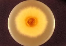
This is a top view of a Sabourauds dextrose agar plate culture of a Microsporum persicolor fungal colony.Created: 1973
-
This is the bottom or reverse view of a Sabourauds dextrose plate culture growing the fungus Microsporum persicolor.Created: 1973
-
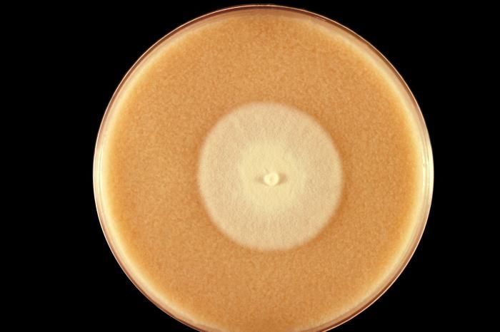
This is a top view of a cereal agar plate culture growing the fungus Microsporum persicolor.Created: 1973
-
This is a top view of a Sabourauds dextrose agar plate culture growing Microsporum persicolor.Created: 1973
-
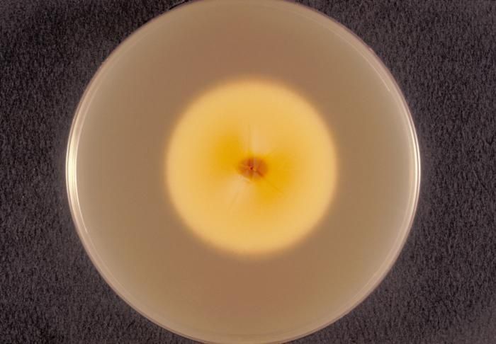
This is the bottom, or reverse view of a Sabourauds dextrose agar plate culture of Microsporum persicolor.Created: 1973
-
This is a top view of a cereal agar plate culture growing a colony of Microsporum persicolor fungus.Created: 1973
-
This is a top view of a Sebourauds dextrose agar plate culture growing a colony of Microsporum persicolor fungus.Created: 1973
-
This is a reverse (bottom) view of a Sebourauds dextrose agar plate culture growing a colony of Microsporum persicolor.Created: 1973
-
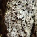
This plate culture is growing the fungus Microsporum persicolor, also known as Trichophyton persicolor.Created: 1964
-
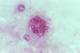
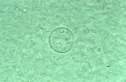
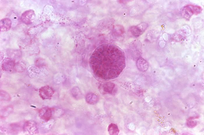
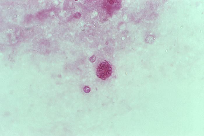
This photomicrograph revealed some of the histopathologic characteristics found within a pus specimen, which was prepared using periodic acid-Schiff (PAS), and which had been harvested from a skin lesion in a case of cutaneous coccidioidomycosis. In this particular specimen youll note the chlamydospore, or immature spherule of a Coccidioides immitis fungal organism. As the reproductive structure of this, as well as other types of fungi, this spherule is also known as a chlamydoconidium, and contains the organisms endospores.Created: 1975
-
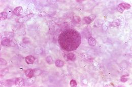
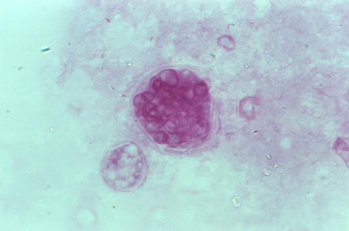
This photomicrograph revealed some of the histopathologic characteristics found within a pus specimen, which was prepared using periodic acid-Schiff (PAS), and which had been harvested from a skin lesion in a case of cutaneous coccidioidomycosis. In this particular specimen youll note the chlamydospore, or mature spherule of a Coccidioides immitis fungal organism. As the reproductive structure of this, as well as other types of fungi, this spherule is also known as a chlamydoconidium, and contains the organisms endospores.Created: 1975
-
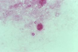
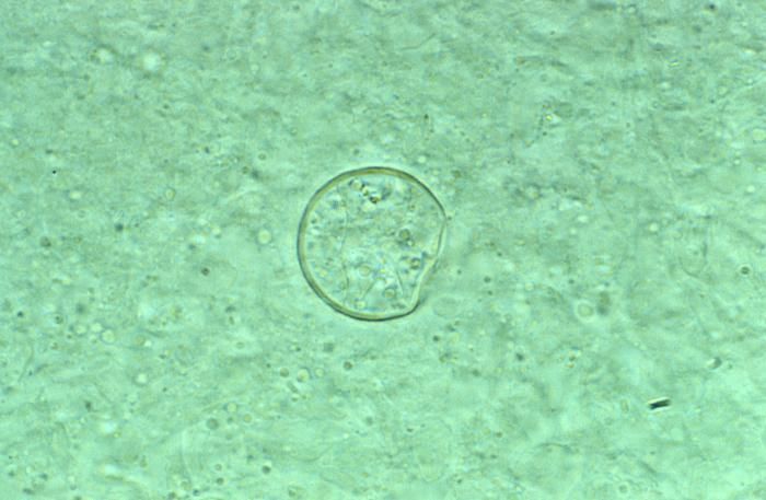
This photomicrograph revealed some of the histopathologic characteristics found within a pus specimen, which was prepared using potassium hudroxide (KOH), and which had been harvested from a skin lesion in a case of cutaneous coccidioidomycosis. In this particular specimen youll note the chlamydospore, or spherule of a Coccidioides immitis fungal organism. As the reproductive structure of this, as well as other types of fungi, this spherule is also known as a chlamydoconidium, and contains the organisms endospores.Created: 1975
-
This was a Sabourauds dextrose agar culture of Coccidioides immitis with chloramphenicol and cycloheximide after 2 wks.Created: 1979