Sivun Microsporum persicolor (Sabour.) Guiart & Grigoraki 1928 kuva
Kuvaus:
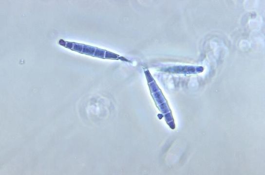
Magnified 500X, this photomicrograph revealed some of the ultrastructural morphology exhibited by the fungal organism, Microsporum persicolor. This particular image highlights details seen in three macroconidia, which are the asexual spores that originate from the filamentous conidiophore. These macroconidia are also known as mitospores, for they are born out of the process of mitosis, and are therefore, haploid when they reach maturity. Unlike the single-celled microconidia, the M. persicolor macroconidia are composed of multiple, attached microconidia, separtated by cell walls, and configured in a cigar-shaped chain.
Created: 1973
Mukana seuraavilla sivuilla:
- Microsporum persicolor
- Ascomycota (kotelosienet)
- Eurotiomycetes
- Onygenales
- Arthrodermataceae
- Microsporum
- Fungi (sienet)
Tämä kuva ei ole esillä missään kokoelmassa.
Lähdetiedot
- lisenssi
- cc-publicdomain
- tarjoaja
- Public Health Image Library
- alkuperäinen
- alkuperäinen mediatiedosto
- käy lähteessä
- kumppanisivusto
- Public Health Image Library
- ID