portrait
Kuvaus:
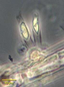
Portrait of colonial form of chrysophyte flagellate, Epipyxis. Cells attach to the base of vase-like loricae by protoplasmic threads containing microtubules. Loricae are constructed of overlapping scales. The scales, visible only by electron-microscopy or staining are composed of interwoven microfibrils. Yellow chloroplast with small stigma (not well-seen in this image). Epipyxis is mixotrophic. Phagotrophy involves bacterial capture by the longer of the two flagella and formation of a feeding "basket" by microtubular action at the anterior of the cell. Often epiphytic on filamentous algae as seen here but sometimes free-swimming. From freshwater pond near Boise, Idaho. Phase contrast.
Mukana seuraavilla sivuilla:
- Life
- Cellular
- Eukaryota (aitotumaiset)
- SAR (Stramenopiles, Alveolates, Rhizaria)
- Stramenopiles (ruskeat levät)
- Ochrophyta
- Chrysophyceae
- Chromulinales
- Dinobryaceae
- Epipyxis
Tämä kuva ei ole esillä missään kokoelmassa.
Lähdetiedot
- lisenssi
- cc-by-nc
- tekijä
- William Bourland
- tarjoaja
- micro*scope
- alkuperäinen
- alkuperäinen mediatiedosto
- käy lähteessä
- kumppanisivusto
- micro*scope
- ID