Brachiomonas simplex Hazen
Kuvaus:
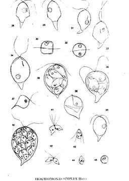
Description: English: Plate 4 Brachiomonas simplex Hazen Drawn 29 June—4 July, 1920, Aalesund, Norway. Figs. 28, 29. Typical mature vegetative cells: in Fig. 29 the stigma lies slightly underneath. Fig. 30. Anterior polar view of similar cell. Fig. 31. Cell on which the "bumps" representing lateral arms are undeveloped. Fig. 32. Anterior polar view of similar cell. Fig. 33. Protoplast ertracted from one of the "bumps" (osmic vapor). Fig. 34. Large older cell with more posterior pyrenoid. Fig. 35 Eight zoospores in motile cell; 12:45 A M' Fig. 36. Four zoosporea in motile cell. Fig. 37. Young zoospore free; chromatophore filling posterior horn. Fig. 38. Three ot the four zoospores have escaped through a triangular rent in the wall of the mother cell. Fig. 39 Similar to Fig. 31, but with more typical position of pyrenoid (osmic vapor) Fig. 40. Thirty-two gametes in motile mother cell; 10 A M. Figs. 41, 42. Gametes at beginning of conjugation (osmic vapor). Fig. 43. Motile zygote, momentarily resting; nuclei not fused. Fig. 44. Zygote, cilia having disappeared, nuclei fused; two stigmata still present. Fig. 45. Zygospore twenty-four hours after conjugation. Date: Published April 1922. (1922-04-DD). Source: Hazen, Tracy E. The phylogeny of the genus Brachiomonas. Bulletin of the Torrey Botanical Club. April, 1922, pp. 87-92. Author: Hazen, Tracy E.
Mukana seuraavilla sivuilla:
- Life
- Cellular
- Eukaryota (aitotumaiset)
- Archaeplastida
- Chloroplastida
- Chlorophyta (Viherlevät)
- Chlorophyceae
- Chlamydomonadales
- Chlamydomonadaceae
- Brachiomonas
Tämä kuva ei ole esillä missään kokoelmassa.
Lähdetiedot
- lisenssi
- cc-publicdomain
- luoja
- Hazen, Tracy E.
- lähde
- Hazen, Tracy E. The phylogeny of the genus Brachiomonas. Bulletin of the Torrey Botanical Club. April, 1922, pp. 87-92.
- alkuperäinen
- alkuperäinen mediatiedosto
- käy lähteessä
- kumppanisivusto
- Wikimedia Commons
- ID