323022 1 En 5 Fig3lr HTML

Kuvaus:
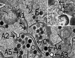
Description: English: Transmission electron micrographs of FHM cells infected by Frog virus 3 (FV3). Enlargement of a viral assembly site showing virions in various stages of assembly. Full (A4 and A5) and empty (A3) viral particles are shown as well as two possible intermediates (A1 and A2) and two aberrant forms (C and E). The inset indicates membranes (arrows), possible originating from the ER, that play a role in virion morphogenesis. Date: 2015. Source: Fig. 3 lower right at https://link.springer.com/chapter/10.1007/978-3-319-13755-1_5 Ranavirus Replication: Molecular, Cellular, and Immunological Events. In: Ranavirus Replication: Molecular, Cellular, and Immunological Events. In: Gray M., Chinchar V. (eds) Ranaviruses. Springer, Cham. doi:10.1007/978-3-319-13755-1_5 . Author: Jancovich J.K., Qin Q., Zhang QY., Chinchar V.G; Gray M., Chinchar V. (eds). Other versions: .
Mukana seuraavilla sivuilla:
Tämä kuva ei ole esillä missään kokoelmassa.
Lähdetiedot
- lisenssi
- cc-by-sa-3.0
- tekijänoikeus
- Jancovich J.K., Qin Q., Zhang QY., Chinchar V.G; Gray M., Chinchar V. (eds)
- luoja
- Jancovich J.K., Qin Q., Zhang QY., Chinchar V.G; Gray M., Chinchar V. (eds)
- lähde
- Fig. 3 lower right at https://link.springer.com/chapter/10.1007/978-3-319-13755-1_5 Ranavirus Replication: Molecular, Cellular, and Immunological Events. In: Ranavirus Replication: Molecular, Cellular, and Immunological Events. In: Gray M., Chinchar V. (eds) Ranaviruses. Springer, Cham. doi:10.1007/978-3-319-13755-1_5
- alkuperäinen
- alkuperäinen mediatiedosto
- käy lähteessä
- kumppanisivusto
- Wikimedia Commons
- ID