Mn5025fig1mag(a)
Kuvaus:
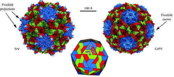
Description: English: Molecular surfaces of Triatoma virus (TrV) and Cricket paralysis virus (CrPV; PDB entry 1b35). The individual proteins are coloured according to the following code: VP1, blue; VP2, green; VP3, red. The structures are on the same scale. TrV displays characteristic surface projections formed by VP1 and VP3 around the fivefold axes, while there is a depression at the twofold axes of TrV. Date: 1 June 2013. Source: https://journals.iucr.org/d/issues/2013/06/00/mn5025/index.html. Author: Gaëlle Squires, Joan Pous, Jon Agirre, Gabriela S. Rozas-Dennis, Marcelo D. Costabel, Gerardo A. Marti, Jorge Navaza, Stéphane Bressanelli, Diego M. A. Guérinb, and Felix A. Reya.
Mukana seuraavilla sivuilla:
Tämä kuva ei ole esillä missään kokoelmassa.
Lähdetiedot
- lisenssi
- cc-by-3.0
- tekijänoikeus
- Gaëlle Squires, Joan Pous, Jon Agirre, Gabriela S. Rozas-Dennis, Marcelo D. Costabel, Gerardo A. Marti, Jorge Navaza, Stéphane Bressanelli, Diego M. A. Guérinb, and Felix A. Reya
- luoja
- Gaëlle Squires, Joan Pous, Jon Agirre, Gabriela S. Rozas-Dennis, Marcelo D. Costabel, Gerardo A. Marti, Jorge Navaza, Stéphane Bressanelli, Diego M. A. Guérinb, and Felix A. Reya
- lähde
- https://journals.iucr.org/d/issues/2013/06/00/mn5025/index.html
- alkuperäinen
- alkuperäinen mediatiedosto
- käy lähteessä
- kumppanisivusto
- Wikimedia Commons
- ID
.jpg)
.jpg)