Pone.0118415.g001A
Kuvaus:
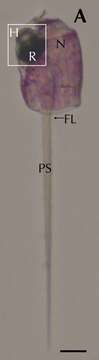
Description: Deutsch: Erythropsidinium spp. and its subcellular structure eyespot “ocelloid”. Light micrographs (LM) of Erythropsidinium spp. H = hyalosome (crystallin body), R = retinal body, N = nucleus, FL = flagella, PS = piston. The ocelloid is located at the left side of a cell seen in ventral view according to the orientation proposed by Kofoid and Swezy. Scale bars: 20 μm. Date: 3 March 2015. Source: Fig. 1A at https://journals.plos.org/plosone/article?id=10.1371/journal.pone.0118415 Function and Evolutionary Origin of Unicellular Camera-Type Eye Structure. PLOS ONE 10(3), PMID 25734540, PMC 4348419, doi:10.1371/journal.pone.0118415 . Author: Shiho Hayakawa, Yasuharu Takaku, Jung Shan Hwang, Takeo Horiguchi, Hiroshi Suga, Walter Gehring, Kazuho Ikeo, Takashi Gojobori. Other versions: .
Mukana seuraavilla sivuilla:
- Life
- Cellular
- Eukaryota (aitotumaiset)
- SAR (Stramenopiles, Alveolates, Rhizaria)
- Alveolata (Alveolaatit)
- Dinophyceae (Panssarilevät)
- Gymnodiniales
- Warnowiaceae
- Erythropsidinium
Tämä kuva ei ole esillä missään kokoelmassa.
Lähdetiedot
- lisenssi
- cc-by-sa-3.0
- tekijänoikeus
- Shiho Hayakawa, Yasuharu Takaku, Jung Shan Hwang, Takeo Horiguchi, Hiroshi Suga, Walter Gehring, Kazuho Ikeo, Takashi Gojobori
- luoja
- Shiho Hayakawa, Yasuharu Takaku, Jung Shan Hwang, Takeo Horiguchi, Hiroshi Suga, Walter Gehring, Kazuho Ikeo, Takashi Gojobori
- lähde
- Fig. 1A at https://journals.plos.org/plosone/article?id=10.1371/journal.pone.0118415 Function and Evolutionary Origin of Unicellular Camera-Type Eye Structure. PLOS ONE 10(3), PMID 25734540, PMC 4348419, doi:10.1371/journal.pone.0118415
- alkuperäinen
- alkuperäinen mediatiedosto
- käy lähteessä
- kumppanisivusto
- Wikimedia Commons
- ID