Sivun Echinoderes hwiizaa Yamasaki & Fujimoto 2014 kuva
Kuvaus:
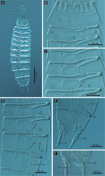
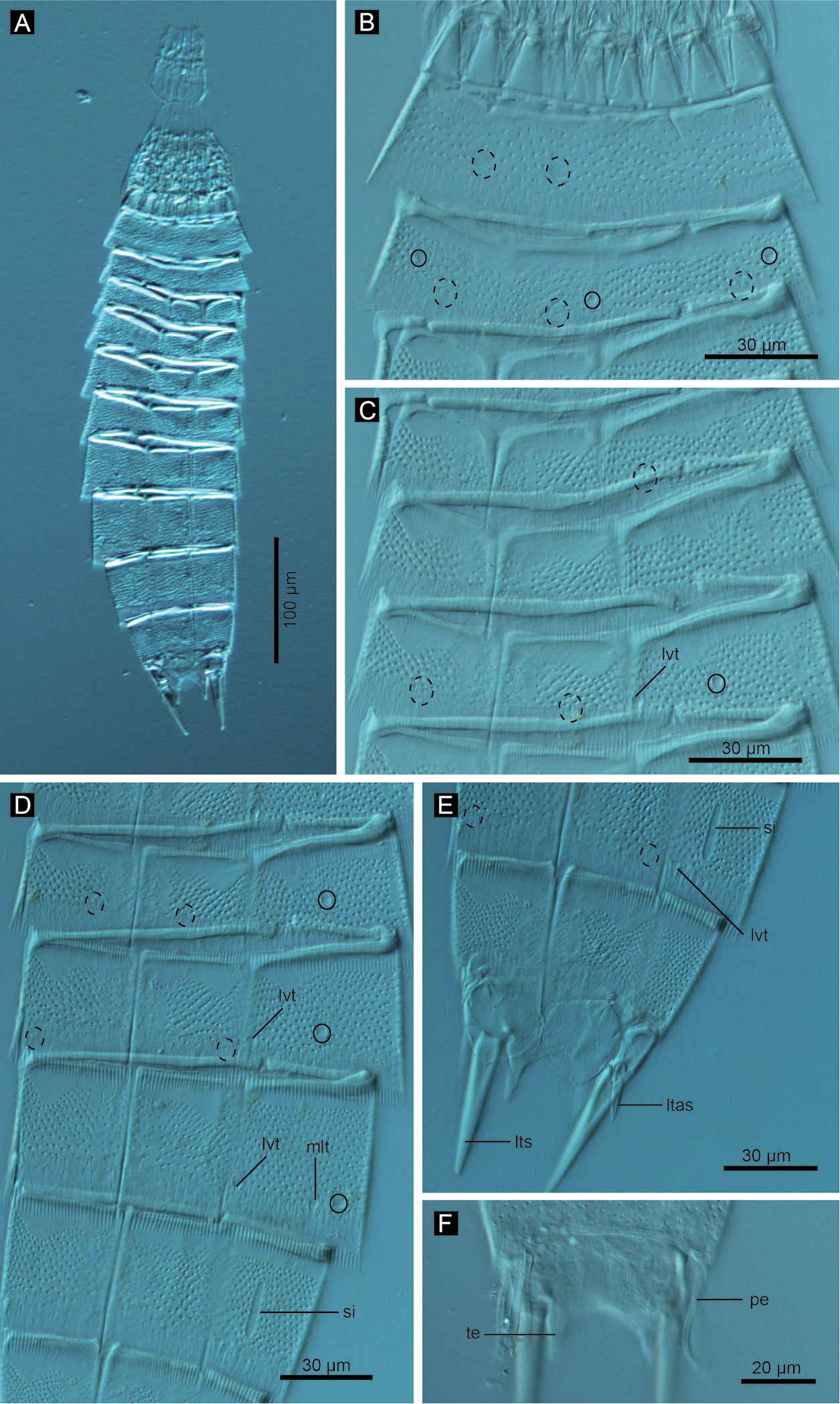
Figure 8.Echinoderes hwiizaa sp. n., Nomarski photomicrographs. A entire animal B segments 1–2, ventral view C segments 3–5, ventral view D segments 6–9 ventral view E segments 9–11 of female, ventral view F segment 11 of male, dorsal view. Complete circles indicate type 2 glandular cell outlet; cashed circles indicate sensory spots. Abbreviations: ltas, lateral terminal accessory spine; lts, lateral terminal spine; lvt, lateroventral tubule; mlt, midlateral tubule; pe, penile spine; si, sieve plate; te, tergal extension.
Mukana seuraavilla sivuilla:
- Life
- Cellular
- Eukaryota (aitotumaiset)
- Opisthokonta
- Metazoa
- Bilateria (Kaksikylkiset)
- Protostomia (Alkusuiset)
- Ecdysozoa
- Kinorhyncha (Okapäämadot)
- Cyclorhagida
- Echinoderidae
- Echinoderes
- Echinoderes hwiizaa
- Scalidophora
Tämä kuva ei ole esillä missään kokoelmassa.
Lähdetiedot
- lisenssi
- cc-by-3.0
- tekijänoikeus
- Hiroshi Yamasaki, Shinta Fujimoto
- bibliografinen lainaus
- Yamasaki H, Fujimoto S (2014) Two new species in the Echinoderes coulli group (Echinoderidae, Cyclorhagida, Kinorhyncha) from the Ryukyu Islands, Japan ZooKeys 382: 27–52
- alkuperäinen
- alkuperäinen mediatiedosto
- käy lähteessä
- kumppanisivusto
- Zookeys
- ID